 Movie
Movie Controller


+ Open data

- Basic information
| Entry | Database: PDB / ID: 7c7c | ||||||
|---|---|---|---|---|---|---|---|
| Title | Crystal structure of human TRAP1 with SJT104 | ||||||
 Components Components | Heat shock protein 75 kDa, mitochondrial Heat shock response Heat shock response | ||||||
 Keywords Keywords | CHAPERONE / TRPA1 / selectivity / mitochondria / Hsp90 / anticancer / drug | ||||||
| Function / homology |  Function and homology information Function and homology informationtranslational attenuation / negative regulation of cellular respiration / Respiratory electron transport / tumor necrosis factor receptor binding / negative regulation of intrinsic apoptotic signaling pathway in response to hydrogen peroxide / chaperone-mediated protein folding / negative regulation of reactive oxygen species biosynthetic process / cell periphery / ATP-dependent protein folding chaperone / mitochondrial intermembrane space ...translational attenuation / negative regulation of cellular respiration / Respiratory electron transport / tumor necrosis factor receptor binding / negative regulation of intrinsic apoptotic signaling pathway in response to hydrogen peroxide / chaperone-mediated protein folding / negative regulation of reactive oxygen species biosynthetic process / cell periphery / ATP-dependent protein folding chaperone / mitochondrial intermembrane space / unfolded protein binding / protein folding / mitochondrial inner membrane / mitochondrial matrix / protein kinase binding / ATP hydrolysis activity / mitochondrion / RNA binding / nucleoplasm / ATP binding / membraneSimilarity search - Function | ||||||
| Biological species |  Homo sapiens (human) Homo sapiens (human) | ||||||
| Method | X-RAY DIFFRACTION / SYNCHROTRON / MOLECULAR REPLACEMENT / Resolution: 3 Å | ||||||
 Authors Authors | Kim, D. / Yang, S. / Yoon, N.G. / Park, E. / Kim, S.Y. / Kang, B.H. / Lee, C. / Kang, S. | ||||||
 Citation Citation | Journal: Acs Med.Chem.Lett. / Year: 2021 Title: Design and Synthesis of TRAP1 Selective Inhibitors: H-Bonding with Asn171 Residue in TRAP1 Increases Paralog Selectivity. Authors: Yang, S. / Yoon, N.G. / Kim, D. / Park, E. / Kim, S.Y. / Lee, J.H. / Lee, C. / Kang, B.H. / Kang, S. | ||||||
| History |
|
- Structure visualization
| Structure viewer | Molecule: MolmilJmol/JSmol |
|---|
- Downloads & links
-Download
| PDBx/mmCIF format | 7c7c.cif.gz | 229.3 KB | Display | PDBx/mmCIF format |
|---|---|---|---|---|
| PDB format | pdb7c7c.ent.gz | 154.6 KB | Display | PDB format |
| PDBx/mmJSON format | 7c7c.json.gz | Tree view | PDBx/mmJSON format | |
| Others |  Other downloads Other downloads |
-Validation report
| Arichive directory | https://data.pdbj.org/pub/pdb/validation_reports/c7/7c7cftp://data.pdbj.org/pub/pdb/validation_reports/c7/7c7c | HTTPS FTP |
|---|
-Related structure data
| Related structure data | 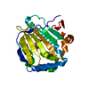 7c7bC 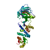 5y3nS S: Starting model for refinement C: citing same article ( |
|---|---|
| Similar structure data |


-Links
 PDBj
PDBj

- Assembly
| Deposited unit | 
| ||||||||||||
|---|---|---|---|---|---|---|---|---|---|---|---|---|---|
| 1 |
| ||||||||||||
| Unit cell |
|
-Components
| #1: Protein | Heat shock response / HSP 75 / TNFR-associated protein 1 / Tumor necrosis factor type 1 receptor-associated protein / TRAP-1 Mass: 57422.090 Da / Num. of mol.: 1 Source method: isolated from a genetically manipulated source Source: (gene. exp.) Homo sapiens (human) / Gene: TRAP1, HSP75 / Production host:  Escherichia coli BL21(DE3) (bacteria) / Strain (production host): BL21(DE3) / References: UniProt: Q12931 Escherichia coli BL21(DE3) (bacteria) / Strain (production host): BL21(DE3) / References: UniProt: Q12931 |
|---|---|
| #2: Chemical | ChemComp-FK0 /   Mass: 372.580 Da / Num. of mol.: 1 / Source method: obtained synthetically / Formula: C12H8BrClFN5O / Feature type: SUBJECT OF INVESTIGATION Mass: 372.580 Da / Num. of mol.: 1 / Source method: obtained synthetically / Formula: C12H8BrClFN5O / Feature type: SUBJECT OF INVESTIGATION |
| Has ligand of interest | Y |
-Experimental details
-Experiment
| Experiment | Method: X-RAY DIFFRACTION / Number of used crystals: 1 |
|---|
- Sample preparation
| Crystal | Density Matthews: 2.65 Å3/Da / Density % sol: 53.51 % |
|---|---|
| Crystal grow | Temperature: 293 K / Method: vapor diffusion, hanging drop / pH: 6.76 Details: 0.1M Cacodylate pH 6.76, 0.1M Calcium acetate, 18% PEG 5000 MME |
-Data collection
| Diffraction | Mean temperature: 100 K / Serial crystal experiment: N |
|---|---|
| Diffraction source | Source: SYNCHROTRON / Site: PAL/PLS  / Beamline: 5C (4A) / Wavelength: 0.9795 Å / Beamline: 5C (4A) / Wavelength: 0.9795 Å |
| Detector | Type: DECTRIS EIGER X 9M / Detector: PIXEL / Date: Mar 22, 2019 |
| Radiation | Protocol: SINGLE WAVELENGTH / Monochromatic (M) / Laue (L): M / Scattering type: x-ray |
| Radiation wavelength | Wavelength: 0.9795 Å / Relative weight: 1 |
| Reflection | Resolution: 3→50 Å / Num. obs: 13023 / % possible obs: 98.8 % / Redundancy: 9.9 % / Biso Wilson estimate: 74.75 Å2 / Rmerge(I) obs: 0.107 / Net I/σ(I): 17.7 |
| Reflection shell | Resolution: 3→3.05 Å / Rmerge(I) obs: 0.52 / Num. unique obs: 13023 |
- Processing
| Software |
| |||||||||||||||||||||||||||||||||||||||||||||||||||||||||||||||||||||||||||
|---|---|---|---|---|---|---|---|---|---|---|---|---|---|---|---|---|---|---|---|---|---|---|---|---|---|---|---|---|---|---|---|---|---|---|---|---|---|---|---|---|---|---|---|---|---|---|---|---|---|---|---|---|---|---|---|---|---|---|---|---|---|---|---|---|---|---|---|---|---|---|---|---|---|---|---|---|
| Refinement | Method to determine structure: MOLECULAR REPLACEMENT Starting model: 5Y3N Resolution: 3→24.98 Å / SU ML: 0.4771 / Cross valid method: FREE R-VALUE / σ(F): 1.44 / Phase error: 28.9653 / Stereochemistry target values: CDL v1.2
| |||||||||||||||||||||||||||||||||||||||||||||||||||||||||||||||||||||||||||
| Solvent computation | Shrinkage radii: 0.9 Å / VDW probe radii: 1.11 Å / Solvent model: FLAT BULK SOLVENT MODEL | |||||||||||||||||||||||||||||||||||||||||||||||||||||||||||||||||||||||||||
| Displacement parameters | Biso mean: 90.32 Å2 | |||||||||||||||||||||||||||||||||||||||||||||||||||||||||||||||||||||||||||
| Refinement step | Cycle: LAST / Resolution: 3→24.98 Å
| |||||||||||||||||||||||||||||||||||||||||||||||||||||||||||||||||||||||||||
| Refine LS restraints |
| |||||||||||||||||||||||||||||||||||||||||||||||||||||||||||||||||||||||||||
| LS refinement shell |
| |||||||||||||||||||||||||||||||||||||||||||||||||||||||||||||||||||||||||||
| Refinement TLS params. | Method: refined / Refine-ID: X-RAY DIFFRACTION
| |||||||||||||||||||||||||||||||||||||||||||||||||||||||||||||||||||||||||||
| Refinement TLS group |
|
