 Movie
Movie Controller

[English] 日本語
 Yorodumi
- PDB-7by9: Malate Dehydrogenase from Geobacillus stearothermophilus (gs-MDH)... -
+ Open data

- Basic information
| Entry | Database: PDB / ID: 7by9 | |||||||||
|---|---|---|---|---|---|---|---|---|---|---|
| Title | Malate Dehydrogenase from Geobacillus stearothermophilus (gs-MDH) complexed with Oxaloacetic Acid (OAA) and Nicotinamide Adenine Dinucleotide (NAD) | |||||||||
 Components Components | Malate dehydrogenase | |||||||||
 Keywords Keywords | OXIDOREDUCTASE / Malate Dehydrogenase | |||||||||
| Function / homology |  Function and homology informationmalate dehydrogenase / L-malate dehydrogenase activity / carboxylic acid metabolic process / tricarboxylic acid cycle Function and homology informationmalate dehydrogenase / L-malate dehydrogenase activity / carboxylic acid metabolic process / tricarboxylic acid cycleSimilarity search - Function | |||||||||
| Biological species |   Geobacillus stearothermophilus (bacteria) Geobacillus stearothermophilus (bacteria) | |||||||||
| Method | X-RAY DIFFRACTION / SYNCHROTRON / MOLECULAR REPLACEMENT / Resolution: 2.2 Å | |||||||||
 Authors Authors | Shimozawa, Y. / Nakamura, T. / Himiyama, T. / Nishiya, Y. | |||||||||
| Funding support |  Japan, 2items Japan, 2items
| |||||||||
 Citation Citation | Journal: J.Biochem. / Year: 2021 Title: Structural analysis and reaction mechanism of malate dehydrogenase from Geobacillus stearothermophilus. Authors: Shimozawa, Y. / Himiyama, T. / Nakamura, T. / Nishiya, Y. | |||||||||
| History |
|
- Structure visualization
| Structure viewer | Molecule: MolmilJmol/JSmol |
|---|
- Downloads & links
-Download
| PDBx/mmCIF format | 7by9.cif.gz | 252.3 KB | Display | PDBx/mmCIF format |
|---|---|---|---|---|
| PDB format | pdb7by9.ent.gz | 197.2 KB | Display | PDB format |
| PDBx/mmJSON format | 7by9.json.gz | Tree view | PDBx/mmJSON format | |
| Others |  Other downloads Other downloads |
-Validation report
| Arichive directory | https://data.pdbj.org/pub/pdb/validation_reports/by/7by9ftp://data.pdbj.org/pub/pdb/validation_reports/by/7by9 | HTTPS FTP |
|---|
-Related structure data
| Related structure data |  7by8SC  7byaC S: Starting model for refinement C: citing same article ( |
|---|---|
| Similar structure data |


-Links
 PDBj
PDBj


- Assembly
| Deposited unit | 
| ||||||||
|---|---|---|---|---|---|---|---|---|---|
| 1 |
| ||||||||
| Unit cell |
|
-Components
| #1: Protein | Mass: 35932.312 Da / Num. of mol.: 4 Source method: isolated from a genetically manipulated source Source: (gene. exp.) Geobacillus stearothermophilus (bacteria)Gene: mdh / Production host: Escherichia coli (E. coli) / References: UniProt: A0A143T1U9, malate dehydrogenase#2: Chemical | ChemComp-NAD / Nicotinamide adenine dinucleotide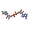  Mass: 663.425 Da / Num. of mol.: 4 / Source method: obtained synthetically / Formula: C21H27N7O14P2 / Feature type: SUBJECT OF INVESTIGATION / Comment: NAD*YM Mass: 663.425 Da / Num. of mol.: 4 / Source method: obtained synthetically / Formula: C21H27N7O14P2 / Feature type: SUBJECT OF INVESTIGATION / Comment: NAD*YM#3: Chemical | ChemComp-OAA / Oxaloacetic acid  Mass: 131.064 Da / Num. of mol.: 4 / Source method: obtained synthetically / Formula: C4H3O5 / Feature type: SUBJECT OF INVESTIGATION Mass: 131.064 Da / Num. of mol.: 4 / Source method: obtained synthetically / Formula: C4H3O5 / Feature type: SUBJECT OF INVESTIGATION#4: Water | ChemComp-HOH / | Water Mass: 18.015 Da / Num. of mol.: 34 / Source method: isolated from a natural source / Formula: H2O Mass: 18.015 Da / Num. of mol.: 34 / Source method: isolated from a natural source / Formula: H2OHas ligand of interest | Y | |
|---|
-Experimental details
-Experiment
| Experiment | Method: X-RAY DIFFRACTION / Number of used crystals: 1 |
|---|
- Sample preparation
| Crystal | Density Matthews: 2.32 Å3/Da / Density % sol: 47.01 % |
|---|---|
| Crystal grow | Temperature: 293 K / Method: vapor diffusion, hanging drop Details: The crystal of wild-type gs-MDH prepared in 0.1 M HEPES (pH 7.5), 10 % polyethylene glycol (PEG) 6000 and 5 % 2-Methyl-2,4-pentanediol (MPD) was soaked into the cryo-protectant containing 2 mM OAA and 2 mM NAD+. |
-Data collection
| Diffraction | Mean temperature: 100 K / Serial crystal experiment: N |
|---|---|
| Diffraction source | Source: SYNCHROTRON / Site: SPring-8 / Beamline: BL26B1 / Wavelength: 1 Å |
| Detector | Type: DECTRIS EIGER X 4M / Detector: PIXEL / Date: Oct 14, 2018 |
| Radiation | Protocol: SINGLE WAVELENGTH / Monochromatic (M) / Laue (L): M / Scattering type: x-ray |
| Radiation wavelength | Wavelength: 1 Å / Relative weight: 1 |
| Reflection | Resolution: 2.2→50 Å / Num. obs: 62899 / % possible obs: 99 % / Redundancy: 4.6 % / CC1/2: 0.966 / Rmerge(I) obs: 0.198 / Net I/σ(I): 4 |
| Reflection shell | Resolution: 2.2→2.26 Å / Rmerge(I) obs: 0.535 / Mean I/σ(I) obs: 1.6 / Num. unique obs: 4635 / CC1/2: 0.913 |
- Processing
| Software |
| |||||||||||||||||||||||||||||||||||||||||||||||||||||||||||||||||||||||||||||||||||||||||||||||||||||||||||||||||||||||||||||||||||||||||||||||||||||||||||
|---|---|---|---|---|---|---|---|---|---|---|---|---|---|---|---|---|---|---|---|---|---|---|---|---|---|---|---|---|---|---|---|---|---|---|---|---|---|---|---|---|---|---|---|---|---|---|---|---|---|---|---|---|---|---|---|---|---|---|---|---|---|---|---|---|---|---|---|---|---|---|---|---|---|---|---|---|---|---|---|---|---|---|---|---|---|---|---|---|---|---|---|---|---|---|---|---|---|---|---|---|---|---|---|---|---|---|---|---|---|---|---|---|---|---|---|---|---|---|---|---|---|---|---|---|---|---|---|---|---|---|---|---|---|---|---|---|---|---|---|---|---|---|---|---|---|---|---|---|---|---|---|---|---|---|---|---|
| Refinement | Method to determine structure: MOLECULAR REPLACEMENT Starting model: 7BY8 Resolution: 2.2→39.393 Å / Cor.coef. Fo:Fc: 0.911 / Cor.coef. Fo:Fc free: 0.866 / SU B: 3.312 / SU ML: 0.096 / Cross valid method: THROUGHOUT / ESU R: 0.336 / ESU R Free: 0.256 Details: Hydrogens have been added in their riding positions
| |||||||||||||||||||||||||||||||||||||||||||||||||||||||||||||||||||||||||||||||||||||||||||||||||||||||||||||||||||||||||||||||||||||||||||||||||||||||||||
| Solvent computation | Ion probe radii: 0.8 Å / Shrinkage radii: 0.8 Å / VDW probe radii: 1.2 Å | |||||||||||||||||||||||||||||||||||||||||||||||||||||||||||||||||||||||||||||||||||||||||||||||||||||||||||||||||||||||||||||||||||||||||||||||||||||||||||
| Displacement parameters | Biso mean: 42.666 Å2
| |||||||||||||||||||||||||||||||||||||||||||||||||||||||||||||||||||||||||||||||||||||||||||||||||||||||||||||||||||||||||||||||||||||||||||||||||||||||||||
| Refinement step | Cycle: LAST / Resolution: 2.2→39.393 Å
| |||||||||||||||||||||||||||||||||||||||||||||||||||||||||||||||||||||||||||||||||||||||||||||||||||||||||||||||||||||||||||||||||||||||||||||||||||||||||||
| Refine LS restraints |
| |||||||||||||||||||||||||||||||||||||||||||||||||||||||||||||||||||||||||||||||||||||||||||||||||||||||||||||||||||||||||||||||||||||||||||||||||||||||||||
| LS refinement shell |
|
