 Movie
Movie Controller


+ Open data

- Basic information
| Entry | Database: PDB / ID: 6zt9 | ||||||
|---|---|---|---|---|---|---|---|
| Title | X-ray structure of mutated arabinofuranosidase | ||||||
 Components Components | Alpha-L-arabinofuranosidase | ||||||
 Keywords Keywords | HYDROLASE / Transferase | ||||||
| Function / homology |  Function and homology information Function and homology informationL-arabinose metabolic process / non-reducing end alpha-L-arabinofuranosidase / alpha-L-arabinofuranosidase activitySimilarity search - Function | ||||||
| Biological species |  Thermobacillus xylanilyticus (bacteria) Thermobacillus xylanilyticus (bacteria) | ||||||
| Method | X-RAY DIFFRACTION / SYNCHROTRON / MOLECULAR REPLACEMENT / Resolution: 2 Å | ||||||
 Authors Authors | Tandrup, T. / Lo Leggio, L. / Zhao, J. / Bissaro, B. / Barbe, S. / Andre, I. / Dumon, C. / O'Donohue, M.J. / Faure, R. | ||||||
 Citation Citation | Journal: N Biotechnol / Year: 2021 Title: Probing the determinants of the transglycosylation/hydrolysis partition in a retaining alpha-l-arabinofuranosidase. Authors: Zhao, J. / Tandrup, T. / Bissaro, B. / Barbe, S. / Poulsen, J.N. / Andre, I. / Dumon, C. / Lo Leggio, L. / O'Donohue, M.J. / Faure, R. | ||||||
| History |
|
- Structure visualization
| Structure viewer | Molecule: MolmilJmol/JSmol |
|---|
- Downloads & links
-Download
| PDBx/mmCIF format | 6zt9.cif.gz | 345.9 KB | Display | PDBx/mmCIF format |
|---|---|---|---|---|
| PDB format | pdb6zt9.ent.gz | 280.2 KB | Display | PDB format |
| PDBx/mmJSON format | 6zt9.json.gz | Tree view | PDBx/mmJSON format | |
| Others |  Other downloads Other downloads |
-Validation report
| Arichive directory | https://data.pdbj.org/pub/pdb/validation_reports/zt/6zt9ftp://data.pdbj.org/pub/pdb/validation_reports/zt/6zt9 | HTTPS FTP |
|---|
-Related structure data
| Related structure data |  6zt6C  6zt7C  6zt8C  6ztaC  2vrqS S: Starting model for refinement C: citing same article ( |
|---|---|
| Similar structure data |


-Links
 PDBj
PDBj- Assembly
| Deposited unit | 
| |||||||||||||||||||||||||||||||||||||||||||||||||||||
|---|---|---|---|---|---|---|---|---|---|---|---|---|---|---|---|---|---|---|---|---|---|---|---|---|---|---|---|---|---|---|---|---|---|---|---|---|---|---|---|---|---|---|---|---|---|---|---|---|---|---|---|---|---|---|
| 1 | 
| |||||||||||||||||||||||||||||||||||||||||||||||||||||
| Unit cell |
| |||||||||||||||||||||||||||||||||||||||||||||||||||||
| Components on special symmetry positions |
| |||||||||||||||||||||||||||||||||||||||||||||||||||||
| Noncrystallographic symmetry (NCS) | NCS domain:
NCS domain segments: Component-ID: 0 / Beg auth comp-ID: VAL / Beg label comp-ID: VAL / End auth comp-ID: GLY / End label comp-ID: GLY / Refine code: 0 / Auth seq-ID: 3 - 496 / Label seq-ID: 3 - 496
NCS ensembles :
|
-Components
-Protein / Sugars , 2 types, 6 molecules ABC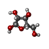
| #1: Protein | Mass: 56227.320 Da / Num. of mol.: 3 Source method: isolated from a genetically manipulated source Source: (gene. exp.) Thermobacillus xylanilyticus (bacteria)Gene: AbjA / Production host: Escherichia coli (E. coli)References: UniProt: O69262, non-reducing end alpha-L-arabinofuranosidase #2: Sugar | Arabinose Type: L-saccharide, alpha linking / Mass: 150.130 Da / Num. of mol.: 3 Type: L-saccharide, alpha linking / Mass: 150.130 Da / Num. of mol.: 3Source method: isolated from a genetically manipulated source Formula: C5H10O5 / Feature type: SUBJECT OF INVESTIGATION |
|---|
-Non-polymers , 4 types, 1321 molecules 
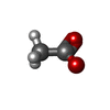


| #3: Chemical | ChemComp-MPD / ( 2-Methyl-2,4-pentanediol Mass: 118.174 Da / Num. of mol.: 21 / Source method: obtained synthetically / Formula: C6H14O2 / Comment: precipitant*YM Mass: 118.174 Da / Num. of mol.: 21 / Source method: obtained synthetically / Formula: C6H14O2 / Comment: precipitant*YM#4: Chemical | ChemComp-ACT / Acetate Mass: 59.044 Da / Num. of mol.: 16 / Source method: obtained synthetically / Formula: C2H3O2 Mass: 59.044 Da / Num. of mol.: 16 / Source method: obtained synthetically / Formula: C2H3O2#5: Chemical | ChemComp-BTB / | Bis-tris methane Mass: 209.240 Da / Num. of mol.: 1 / Source method: obtained synthetically / Formula: C8H19NO5 / Comment: pH buffer*YM Mass: 209.240 Da / Num. of mol.: 1 / Source method: obtained synthetically / Formula: C8H19NO5 / Comment: pH buffer*YM#6: Water | ChemComp-HOH / | WaterMass: 18.015 Da / Num. of mol.: 1283 / Source method: isolated from a natural source / Formula: H2O |
|---|
-Details
| Has ligand of interest | Y |
|---|
-Experimental details
-Experiment
| Experiment | Method: X-RAY DIFFRACTION / Number of used crystals: 1 |
|---|
- Sample preparation
| Crystal | Density Matthews: 4.01 Å3/Da / Density % sol: 69.3 % |
|---|---|
| Crystal grow | Temperature: 298 K / Method: vapor diffusion, sitting drop / pH: 5.5 Details: 0.2 M ammonium acetate, 0.1 M Bis-tris pH 5.5, 45% (v/v) MPD |
-Data collection
| Diffraction | Mean temperature: 100 K / Serial crystal experiment: N |
|---|---|
| Diffraction source | Source: SYNCHROTRON / Site: ESRF  / Beamline: ID30B / Wavelength: 0.969 Å / Beamline: ID30B / Wavelength: 0.969 Å |
| Detector | Type: DECTRIS PILATUS 6M / Detector: PIXEL / Date: Feb 12, 2016 |
| Radiation | Protocol: SINGLE WAVELENGTH / Monochromatic (M) / Laue (L): M / Scattering type: x-ray |
| Radiation wavelength | Wavelength: 0.969 Å / Relative weight: 1 |
| Reflection | Resolution: 2→136.02 Å / Num. obs: 185418 / % possible obs: 99.9 % / Redundancy: 21.71 % / CC1/2: 0.998 / Net I/σ(I): 11.19 |
| Reflection shell | Resolution: 2→2.05 Å / Num. unique obs: 13515 / CC1/2: 0.503 |
- Processing
| Software |
| ||||||||||||||||||||||||||||||||||||||||||||||||||||||||||||
|---|---|---|---|---|---|---|---|---|---|---|---|---|---|---|---|---|---|---|---|---|---|---|---|---|---|---|---|---|---|---|---|---|---|---|---|---|---|---|---|---|---|---|---|---|---|---|---|---|---|---|---|---|---|---|---|---|---|---|---|---|---|
| Refinement | Method to determine structure: MOLECULAR REPLACEMENT Starting model: 2VRQ Resolution: 2→136.02 Å / Cor.coef. Fo:Fc: 0.977 / Cor.coef. Fo:Fc free: 0.969 / SU B: 3.243 / SU ML: 0.081 / Cross valid method: THROUGHOUT / σ(F): 0 / ESU R: 0.096 / ESU R Free: 0.095 / Stereochemistry target values: MAXIMUM LIKELIHOOD Details: HYDROGENS HAVE BEEN ADDED IN THE RIDING POSITIONS U VALUES : REFINED INDIVIDUALLY
| ||||||||||||||||||||||||||||||||||||||||||||||||||||||||||||
| Solvent computation | Ion probe radii: 0.8 Å / Shrinkage radii: 0.8 Å / VDW probe radii: 1.2 Å / Solvent model: MASK | ||||||||||||||||||||||||||||||||||||||||||||||||||||||||||||
| Displacement parameters | Biso max: 150.14 Å2 / Biso mean: 40.186 Å2 / Biso min: 19.23 Å2
| ||||||||||||||||||||||||||||||||||||||||||||||||||||||||||||
| Refinement step | Cycle: final / Resolution: 2→136.02 Å
| ||||||||||||||||||||||||||||||||||||||||||||||||||||||||||||
| Refine LS restraints |
| ||||||||||||||||||||||||||||||||||||||||||||||||||||||||||||
| Refine LS restraints NCS | Refine-ID: X-RAY DIFFRACTION / Type: interatomic distance / Rms dev position: 0.06 Å / Weight position: 0.05
| ||||||||||||||||||||||||||||||||||||||||||||||||||||||||||||
| LS refinement shell | Resolution: 2→2.052 Å / Rfactor Rfree error: 0 / Total num. of bins used: 20
|
