[English] 日本語
Yorodumi
- PDB-6u91: Crystal structure of DNMT3B(Q772R)-DNMT3L in complex with CpGpT DNA -

+
Open data


ID or keywords:

Loading...

-
Basic information

Entry
Database: PDB / ID: 6u91
TitleCrystal structure of DNMT3B(Q772R)-DNMT3L in complex with CpGpT DNA
Components
  • (DNA (cytosine-5)-methyltransferase ...) x 2
  • CpGpT DNA (25-MER)
KeywordsTransferase/DNA / DNMT3B / DNMT3L / DNA methylation / Methyltransferase / transferase-DNA complex / TRANSFERASE
Function / homology
Function and homology information


retrotransposon silencing by heterochromatin formation / : / epigenetic programing of female pronucleus / chorionic trophoblast cell differentiation / DNA (cytosine-5-)-methyltransferase activity, acting on CpG substrates / DNA-methyltransferase activity / genomic imprinting / DNA (cytosine-5-)-methyltransferase / DNA (cytosine-5-)-methyltransferase activity / autosome genomic imprinting ...retrotransposon silencing by heterochromatin formation / : / epigenetic programing of female pronucleus / chorionic trophoblast cell differentiation / DNA (cytosine-5-)-methyltransferase activity, acting on CpG substrates / DNA-methyltransferase activity / genomic imprinting / DNA (cytosine-5-)-methyltransferase / DNA (cytosine-5-)-methyltransferase activity / autosome genomic imprinting / negative regulation of DNA methylation-dependent heterochromatin formation / SUMOylation of DNA methylation proteins / DNA methylation-dependent heterochromatin formation / ESC/E(Z) complex / : / negative regulation of gene expression, epigenetic / DNA metabolic process / male meiosis I / catalytic complex / heterochromatin / enzyme activator activity / DNA methylation / placenta development / PRC2 methylates histones and DNA / condensed nuclear chromosome / Defective pyroptosis / stem cell differentiation / NoRC negatively regulates rRNA expression / transcription corepressor activity / methylation / spermatogenesis / positive regulation of gene expression / negative regulation of transcription by RNA polymerase II / enzyme binding / DNA binding / nucleoplasm / metal ion binding / nucleus / cytosol
Similarity search - Function
DNA (cytosine-5)-methyltransferase 3B, ADD domain / : / DNMT3, ADD PHD zinc finger / DNMT3, cysteine rich ADD domain / DNMT3, cysteine rich ADD domain, GATA1-like zinc finger / ADD domain / ADD domain profile. / DNA methylase, C-5 cytosine-specific, active site / C-5 cytosine-specific DNA methylases active site. / C-5 cytosine-specific DNA methylase (Dnmt) domain profile. ...DNA (cytosine-5)-methyltransferase 3B, ADD domain / : / DNMT3, ADD PHD zinc finger / DNMT3, cysteine rich ADD domain / DNMT3, cysteine rich ADD domain, GATA1-like zinc finger / ADD domain / ADD domain profile. / DNA methylase, C-5 cytosine-specific, active site / C-5 cytosine-specific DNA methylases active site. / C-5 cytosine-specific DNA methylase (Dnmt) domain profile. / C-5 cytosine methyltransferase / C-5 cytosine-specific DNA methylase / domain with conserved PWWP motif / PWWP domain / PWWP domain profile. / PWWP domain / Vaccinia Virus protein VP39 / Zinc finger, FYVE/PHD-type / Zinc finger, RING/FYVE/PHD-type / S-adenosyl-L-methionine-dependent methyltransferase superfamily / Rossmann fold / 3-Layer(aba) Sandwich / Alpha Beta
Similarity search - Domain/homology
S-ADENOSYL-L-HOMOCYSTEINE / DNA / DNA (> 10) / DNA (cytosine-5)-methyltransferase 3B / DNA (cytosine-5)-methyltransferase 3-like
Similarity search - Component
Biological speciesHomo sapiens (human)
MethodX-RAY DIFFRACTION / SYNCHROTRON / MOLECULAR REPLACEMENT / Resolution: 2.99998872297 Å
AuthorsGao, L. / Song, J.
Funding support United States, 1items
OrganizationGrant numberCountry
National Institutes of Health/National Human Genome Research Institute (NIH/NHGRI)1R35GM119721 United States
Citation
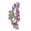
Journal: To Be Published
Title: Comprehensive structure-function characterization of DNMT3B and DNMT3A reveals distinctive de novo DNA methylation mechanisms
Authors: Gao, L.
History
DepositionSep 6, 2019Deposition site: RCSB / Processing site: RCSB
Revision 1.0Jun 10, 2020Provider: repository / Type: Initial release
Revision 1.1Oct 11, 2023Group: Data collection / Database references ...Data collection / Database references / Derived calculations / Refinement description
Category: chem_comp_atom / chem_comp_bond ...chem_comp_atom / chem_comp_bond / database_2 / pdbx_initial_refinement_model / struct_conn / struct_conn_type
Item: _database_2.pdbx_DOI / _database_2.pdbx_database_accession ..._database_2.pdbx_DOI / _database_2.pdbx_database_accession / _struct_conn.conn_type_id / _struct_conn.id / _struct_conn.pdbx_dist_value / _struct_conn.pdbx_leaving_atom_flag / _struct_conn.ptnr1_auth_asym_id / _struct_conn.ptnr1_auth_comp_id / _struct_conn.ptnr1_auth_seq_id / _struct_conn.ptnr1_label_asym_id / _struct_conn.ptnr1_label_atom_id / _struct_conn.ptnr1_label_comp_id / _struct_conn.ptnr1_label_seq_id / _struct_conn.ptnr2_auth_asym_id / _struct_conn.ptnr2_auth_comp_id / _struct_conn.ptnr2_auth_seq_id / _struct_conn.ptnr2_label_asym_id / _struct_conn.ptnr2_label_atom_id / _struct_conn.ptnr2_label_comp_id / _struct_conn.ptnr2_label_seq_id / _struct_conn_type.id

-
Structure visualization

Structure viewerMolecule:
MolmilJmol/JSmol

Downloads & links

-
Assembly

Deposited unit
A: DNA (cytosine-5)-methyltransferase 3B
B: DNA (cytosine-5)-methyltransferase 3-like
C: DNA (cytosine-5)-methyltransferase 3-like
D: DNA (cytosine-5)-methyltransferase 3B
E: CpGpT DNA (25-MER)
F: CpGpT DNA (25-MER)
hetero molecules


Theoretical massNumber of molelcules
Total (without water)130,99811
Polymers130,1566
Non-polymers8425
Water57632
1


  • Idetical with deposited unit
  • defined by author
  • Evidence: gel filtration
TypeNameSymmetry operationNumber
identity operation1_555x,y,z1
Unit cell
Length a, b, c (Å)190.344, 190.344, 49.644
Angle α, β, γ (deg.)90.000, 90.000, 120.000
Int Tables number144
Space group name H-MP31
Space group name HallP31

-
Components

-
DNA (cytosine-5)-methyltransferase ... , 2 types, 4 molecules ADBC

#1: Protein DNA (cytosine-5)-methyltransferase 3B / Dnmt3b / DNA methyltransferase HsaIIIB / M.HsaIIIB


Mass: 33215.477 Da / Num. of mol.: 2 / Mutation: Q772R
Source method: isolated from a genetically manipulated source
Source: (gene. exp.) Homo sapiens (human) / Gene: DNMT3B / Production host: Escherichia coli (E. coli) / Variant (production host): DE3
References: UniProt: Q9UBC3, DNA (cytosine-5-)-methyltransferase
#2: Protein DNA (cytosine-5)-methyltransferase 3-like


Mass: 24163.705 Da / Num. of mol.: 2
Source method: isolated from a genetically manipulated source
Details: DNA (cytosine-5)-methyltransferase 3-like / Source: (gene. exp.) Homo sapiens (human) / Gene: DNMT3L / Production host: Escherichia coli (E. coli) / Variant (production host): DE3 / References: UniProt: Q9UJW3

-
DNA chain , 1 types, 2 molecules EF

#3: DNA chain CpGpT DNA (25-MER)


Mass: 7698.969 Da / Num. of mol.: 2 / Source method: obtained synthetically / Source: (synth.) Homo sapiens (human)

-
Non-polymers , 3 types, 37 molecules

#4: Chemical ChemComp-MG / MAGNESIUM ION


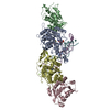
Mass: 24.305 Da / Num. of mol.: 3 / Source method: obtained synthetically / Formula: Mg
#5: Chemical ChemComp-SAH / S-ADENOSYL-L-HOMOCYSTEINE / S-Adenosyl-L-homocysteine


Type: L-peptide linking / Mass: 384.411 Da / Num. of mol.: 2 / Source method: obtained synthetically / Formula: C14H20N6O5S / Feature type: SUBJECT OF INVESTIGATION
#6: Water ChemComp-HOH / water / Water


Mass: 18.015 Da / Num. of mol.: 32 / Source method: isolated from a natural source / Formula: H2O

-
Details

Has ligand of interestY

-
Experimental details

-
Experiment

ExperimentMethod: X-RAY DIFFRACTION / Number of used crystals: 1

-
Sample preparation

CrystalDensity Matthews: 3.99 Å3/Da / Density % sol: 69.17 %
Crystal growTemperature: 277.15 K / Method: vapor diffusion, hanging drop / pH: 8 / Details: 0.1 M Tris-HCl (pH 8.0), 200 mM MgCl2, 8% PEG 4000

-
Data collection

DiffractionMean temperature: 100 K / Serial crystal experiment: N
Diffraction sourceSource: SYNCHROTRON / Site: ALS / Beamline: 5.0.1 / Wavelength: 0.9774 Å
DetectorType: ADSC QUANTUM 315r / Detector: CCD / Date: Mar 7, 2018
RadiationProtocol: SINGLE WAVELENGTH / Monochromatic (M) / Laue (L): M / Scattering type: x-ray
Radiation wavelengthWavelength: 0.9774 Å / Relative weight: 1
ReflectionResolution: 2.99998872297→47.59 Å / Num. obs: 40256 / % possible obs: 99.7 % / Redundancy: 8.2 % / Biso Wilson estimate: 64.1976072599 Å2 / CC1/2: 0.99 / Net I/σ(I): 6.76
Reflection shellResolution: 3→3.11 Å / Redundancy: 6.8 % / Rmerge(I) obs: 1.36 / Num. unique obs: 4048 / CC1/2: 0.704 / % possible all: 98.1

-
Processing

Software
NameVersionClassification
PHENIX1.12_2829refinement
HKL-2000data reduction
HKL-2000data scaling
PHASERphasing
RefinementMethod to determine structure: MOLECULAR REPLACEMENT
Starting model: 5YX2
Resolution: 2.99998872297→47.586 Å / SU ML: 0.367122545453 / Cross valid method: FREE R-VALUE / σ(F): 1.95924514285 / Phase error: 27.4475126822
RfactorNum. reflection% reflection
Rfree0.25910047627 2018 5.02252420419 %
Rwork0.214549750174 --
obs0.216782524271 40179 99.6948042281 %
Solvent computationShrinkage radii: 0.9 Å / VDW probe radii: 1.11 Å
Displacement parametersBiso mean: 75.9153995093 Å2
Refinement stepCycle: LAST / Resolution: 2.99998872297→47.586 Å
ProteinNucleic acidLigandSolventTotal
Num. atoms7493 1020 55 32 8600
Refine LS restraints
Refine-IDTypeDev idealNumber
X-RAY DIFFRACTIONf_bond_d0.004292201539378925
X-RAY DIFFRACTIONf_angle_d0.65271053563112350
X-RAY DIFFRACTIONf_chiral_restr0.04176685233311347
X-RAY DIFFRACTIONf_plane_restr0.003934915024711422
X-RAY DIFFRACTIONf_dihedral_angle_d21.39292019315078
LS refinement shell
Resolution (Å)Rfactor RfreeNum. reflection RfreeRfactor RworkNum. reflection RworkRefine-ID% reflection obs (%)
3-3.0750.3849488320471400.3380456847092738X-RAY DIFFRACTION97.957794418
3.075-3.15810.3242604349841440.3039117869522670X-RAY DIFFRACTION99.1194082423
3.1581-3.2510.348993234031270.3039868383662729X-RAY DIFFRACTION99.7555012225
3.251-3.35590.3592913388311380.2859095476412758X-RAY DIFFRACTION99.89651604
3.3559-3.47590.3138953048921440.2531435207282709X-RAY DIFFRACTION99.8599929996
3.4759-3.6150.2826213808611460.2332525968852731X-RAY DIFFRACTION100
3.615-3.77940.2307102836151480.2088753583432765X-RAY DIFFRACTION99.9313893654
3.7794-3.97860.238722393111460.202430275552701X-RAY DIFFRACTION99.9648876404
3.9786-4.22770.264728574131560.1898961923062732X-RAY DIFFRACTION100
4.2277-4.55390.202341576771440.1688637385862730X-RAY DIFFRACTION99.9304589708
4.5539-5.01170.2122637588661480.172358630512734X-RAY DIFFRACTION99.9306518724
5.0117-5.73590.2636786602671350.1975313689372727X-RAY DIFFRACTION99.9650716032
5.7359-7.22260.2165313344721500.2151365394342726X-RAY DIFFRACTION99.8957971518
7.2226-47.5860.2559259992421520.1963533989332711X-RAY DIFFRACTION99.5826086957

+
About Yorodumi

-
News

-
Feb 9, 2022. New format data for meta-information of EMDB entries

New format data for meta-information of EMDB entries

  • Version 3 of the EMDB header file is now the official format.
  • The previous official version 1.9 will be removed from the archive.

Related info.:EMDB header

External links:wwPDB to switch to version 3 of the EMDB data model

-
Aug 12, 2020. Covid-19 info

Covid-19 info

URL: https://pdbj.org/emnavi/covid19.php

New page: Covid-19 featured information page in EM Navigator.

Related info.:Covid-19 info / Mar 5, 2020. Novel coronavirus structure data

+
Mar 5, 2020. Novel coronavirus structure data

Novel coronavirus structure data

Related info.:Yorodumi Speices / Aug 12, 2020. Covid-19 info

External links:COVID-19 featured content - PDBj / Molecule of the Month (242):Coronavirus Proteases

+
Jan 31, 2019. EMDB accession codes are about to change! (news from PDBe EMDB page)

EMDB accession codes are about to change! (news from PDBe EMDB page)

  • The allocation of 4 digits for EMDB accession codes will soon come to an end. Whilst these codes will remain in use, new EMDB accession codes will include an additional digit and will expand incrementally as the available range of codes is exhausted. The current 4-digit format prefixed with “EMD-” (i.e. EMD-XXXX) will advance to a 5-digit format (i.e. EMD-XXXXX), and so on. It is currently estimated that the 4-digit codes will be depleted around Spring 2019, at which point the 5-digit format will come into force.
  • The EM Navigator/Yorodumi systems omit the EMD- prefix.

Related info.:Q: What is EMD? / ID/Accession-code notation in Yorodumi/EM Navigator

External links:EMDB Accession Codes are Changing Soon! / Contact to PDBj

+
Jul 12, 2017. Major update of PDB

Major update of PDB

  • wwPDB released updated PDB data conforming to the new PDBx/mmCIF dictionary.
  • This is a major update changing the version number from 4 to 5, and with Remediation, in which all the entries are updated.
  • In this update, many items about electron microscopy experimental information are reorganized (e.g. em_software).
  • Now, EM Navigator and Yorodumi are based on the updated data.

External links:wwPDB Remediation / Enriched Model Files Conforming to OneDep Data Standards Now Available in the PDB FTP Archive

-
Yorodumi

Thousand views of thousand structures

  • Yorodumi is a browser for structure data from EMDB, PDB, SASBDB, etc.
  • This page is also the successor to EM Navigator detail page, and also detail information page/front-end page for Omokage search.
  • The word "yorodu" (or yorozu) is an old Japanese word meaning "ten thousand". "mi" (miru) is to see.

Related info.:EMDB / PDB / SASBDB / Comparison of 3 databanks / Yorodumi Search / Aug 31, 2016. New EM Navigator & Yorodumi / Yorodumi Papers / Jmol/JSmol / Function and homology information / Changes in new EM Navigator and Yorodumi

Read more