 Movie
Movie Controller

[English] 日本語
 Yorodumi
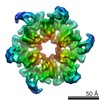
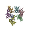
- PDB-6rhw: Crystal structure of human CD11b I-domain (CD11b-I) in complex wi... -
+ Open data

- Basic information
| Entry | Database: PDB / ID: 6rhw | ||||||
|---|---|---|---|---|---|---|---|
| Title | Crystal structure of human CD11b I-domain (CD11b-I) in complex with Staphylococcus aureus octameric bi-component leukocidin LukGH | ||||||
 Components Components |
| ||||||
 Keywords Keywords |  TOXIN / LEUKOCIDIN / PORE-FORMING TOXIN / OCTAMER / receptor / complex TOXIN / LEUKOCIDIN / PORE-FORMING TOXIN / OCTAMER / receptor / complex | ||||||
| Function / homology |  Function and homology information Function and homology informationectodermal cell differentiation / integrin alphaM-beta2 complex / positive regulation of prostaglandin-E synthase activity / response to curcumin / positive regulation of neutrophil degranulation / response to Gram-positive bacterium / positive regulation of microglial cell mediated cytotoxicity / complement component C3b binding / cytolysis in another organism / vertebrate eye-specific patterning ...ectodermal cell differentiation / integrin alphaM-beta2 complex / positive regulation of prostaglandin-E synthase activity / response to curcumin / positive regulation of neutrophil degranulation / response to Gram-positive bacterium / positive regulation of microglial cell mediated cytotoxicity / complement component C3b binding / cytolysis in another organism / vertebrate eye-specific patterning / complement-mediated synapse pruning / Toll Like Receptor 4 (TLR4) Cascade / negative regulation of dopamine metabolic process / cell-cell adhesion via plasma-membrane adhesion molecules / complement receptor mediated signaling pathway / heterotypic cell-cell adhesion / integrin complex / cargo receptor activity / cell adhesion mediated by integrin / phagocytosis, engulfment / amyloid-beta clearance / plasma membrane raft / tertiary granule membrane / positive regulation of protein targeting to membrane / Integrin cell surface interactions / specific granule membrane / response to mechanical stimulus / forebrain development / heat shock protein binding / receptor-mediated endocytosis / cell-matrix adhesion / positive regulation of superoxide anion generation / integrin-mediated signaling pathway / response to ischemia / Cell surface interactions at the vascular wall / microglial cell activation / cell-cell adhesion / integrin binding / response to estradiol / amyloid-beta binding / Interleukin-4 and Interleukin-13 signaling / cell adhesion / external side of plasma membrane / innate immune response / Neutrophil degranulation / cell surface / extracellular space / extracellular exosome / extracellular region / metal ion binding / plasma membraneSimilarity search - Function | ||||||
| Biological species |   Staphylococcus aureus (bacteria) Staphylococcus aureus (bacteria) Homo sapiens (human) Homo sapiens (human) | ||||||
| Method | X-RAY DIFFRACTION / SYNCHROTRON / MOLECULAR REPLACEMENT / Resolution: 2.75 Å | ||||||
 Authors Authors | Trstenjak, N. / Milic, D. / Djinovic-Carugo, K. / Badarau, A. | ||||||
| Funding support |  Austria, 1items Austria, 1items
| ||||||
 Citation Citation | Journal: Proc.Natl.Acad.Sci.USA / Year: 2020 Title: Molecular mechanism of leukocidin GH-integrin CD11b/CD18 recognition and species specificity. Authors: Trstenjak, N. / Milic, D. / Graewert, M.A. / Rouha, H. / Svergun, D. / Djinovic-Carugo, K. / Nagy, E. / Badarau, A. | ||||||
| History |
|
- Structure visualization
| Structure viewer | Molecule: MolmilJmol/JSmol |
|---|
- Downloads & links
-Download
| PDBx/mmCIF format | 6rhw.cif.gz | 509 KB | Display | PDBx/mmCIF format |
|---|---|---|---|---|
| PDB format | pdb6rhw.ent.gz | 351.7 KB | Display | PDB format |
| PDBx/mmJSON format | 6rhw.json.gz | Tree view | PDBx/mmJSON format | |
| Others |  Other downloads Other downloads |
-Validation report
| Arichive directory | https://data.pdbj.org/pub/pdb/validation_reports/rh/6rhwftp://data.pdbj.org/pub/pdb/validation_reports/rh/6rhw | HTTPS FTP |
|---|
-Related structure data
| Related structure data |  6rhvC  1idoS S: Starting model for refinement C: citing same article ( |
|---|---|
| Similar structure data |


-Links
 PDBj
PDBj


- Assembly
| Deposited unit | 
| ||||||||||||
|---|---|---|---|---|---|---|---|---|---|---|---|---|---|
| 1 | 
| ||||||||||||
| Unit cell |
|
-Components
-Beta-channel forming ... , 2 types, 2 molecules GH
| #1: Protein | Mass: 35625.309 Da / Num. of mol.: 1 Source method: isolated from a genetically manipulated source Source: (gene. exp.) Staphylococcus aureus (bacteria)Gene: lukG, ELQ85_15505, EP54_11070, EQ90_09460, HMPREF3211_02235, NCTC10654_02179, NCTC10702_03203, NCTC13131_01350, RK64_10675 Production host: Escherichia coli BL21(DE3) (bacteria) / Variant (production host): TUNER / References: UniProt: A0A0D6HCK9, UniProt: Q2FWP0*PLUS |
|---|---|
| #2: Protein | Mass: 37682.680 Da / Num. of mol.: 1 Source method: isolated from a genetically manipulated source Source: (gene. exp.) Staphylococcus aureus (bacteria)Gene: lukH, BTN44_11630, EP54_11065, EQ90_09455, HMPREF3211_02234, NCTC10654_02180, NCTC10702_03204, NCTC13131_01351, NCTC13196_01958, RK64_10680 Production host: Escherichia coli BL21(DE3) (bacteria) / Variant (production host): TUNER / References: UniProt: A0A0D6HC73, UniProt: Q2FWN9*PLUS |
-Protein , 1 types, 1 molecules C
| #3: Protein | Mass: 22293.439 Da / Num. of mol.: 1 Source method: isolated from a genetically manipulated source Source: (gene. exp.) Homo sapiens (human) / Gene: ITGAM, CD11B, CR3A / Production host: Escherichia coli BL21(DE3) (bacteria) / Variant (production host): TUNER / References: UniProt: P11215 |
|---|
-Non-polymers , 3 types, 9 molecules 


| #4: Chemical | ChemComp-DMS / Dimethyl sulfoxide Mass: 78.133 Da / Num. of mol.: 6 / Source method: obtained synthetically / Formula: C2H6OS / Comment: DMSO, precipitant*YM Mass: 78.133 Da / Num. of mol.: 6 / Source method: obtained synthetically / Formula: C2H6OS / Comment: DMSO, precipitant*YM#5: Chemical | ChemComp-MG / |  Mass: 24.305 Da / Num. of mol.: 1 / Source method: obtained synthetically / Formula: Mg Mass: 24.305 Da / Num. of mol.: 1 / Source method: obtained synthetically / Formula: Mg#6: Water | ChemComp-HOH / | WaterMass: 18.015 Da / Num. of mol.: 2 / Source method: isolated from a natural source / Formula: H2O |
|---|
-Experimental details
-Experiment
| Experiment | Method: X-RAY DIFFRACTION / Number of used crystals: 1 |
|---|
- Sample preparation
| Crystal | Density Matthews: 2.61 Å3/Da / Density % sol: 52.81 % |
|---|---|
| Crystal grow | Temperature: 295 K / Method: vapor diffusion, hanging drop / pH: 7.5 Details: Crystallization drops were prepared by mixing 1.0 uL LukGH/huCD11b-I complex (5.2 mg/mL) in 25 mM HEPES (pH 7.5), 1 mM MgCl2 with 0.5 uL reservoir solution containing 30% (v/v) Jeffamine-600 and 10% (v/v) DMSO. |
-Data collection
| Diffraction | Mean temperature: 100 K / Serial crystal experiment: N |
|---|---|
| Diffraction source | Source: SYNCHROTRON / Site: ESRF  / Beamline: ID29 / Wavelength: 1.07227 Å / Beamline: ID29 / Wavelength: 1.07227 Å |
| Detector | Type: DECTRIS PILATUS 6M / Detector: PIXEL / Date: Nov 22, 2018 |
| Radiation | Protocol: SINGLE WAVELENGTH / Monochromatic (M) / Laue (L): M / Scattering type: x-ray |
| Radiation wavelength | Wavelength: 1.07227 Å / Relative weight: 1 |
| Reflection | Resolution: 2.748→45.081 Å / Num. obs: 14539 / % possible obs: 53.8 % / Redundancy: 12.6 % / Biso Wilson estimate: 53.18 Å2 / CC1/2: 0.996 / Rmerge(I) obs: 0.31 / Rpim(I) all: 0.09 / Rrim(I) all: 0.347 / Net I/σ(I): 7.8 |
| Reflection shell | Resolution: 2.748→2.994 Å / Redundancy: 12.5 % / Rmerge(I) obs: 1.668 / Mean I/σ(I) obs: 1.8 / Num. unique obs: 902 / CC1/2: 0.627 / Rpim(I) all: 0.486 / Rrim(I) all: 1.739 / % possible all: 15 |
- Processing
| Software |
| ||||||||||||||||||||||||||||||||||||||||||||||||||||||||||||||||||||||||||||||||||||||||||||||||||||
|---|---|---|---|---|---|---|---|---|---|---|---|---|---|---|---|---|---|---|---|---|---|---|---|---|---|---|---|---|---|---|---|---|---|---|---|---|---|---|---|---|---|---|---|---|---|---|---|---|---|---|---|---|---|---|---|---|---|---|---|---|---|---|---|---|---|---|---|---|---|---|---|---|---|---|---|---|---|---|---|---|---|---|---|---|---|---|---|---|---|---|---|---|---|---|---|---|---|---|---|---|---|
| Refinement | Method to determine structure: MOLECULAR REPLACEMENT Starting model: LukGH dimer from the structure of its complex with mouse CD11b I-domain (CD11b-I) and the modified huCD11b-I domain (PDB code 1IDO) lacking the C-terminal alpha-helix (residues 303-315) Resolution: 2.75→45.08 Å / SU ML: 0.3789 / Cross valid method: FREE R-VALUE / σ(F): 1.33 / Phase error: 30.8887 Details: The structure was refined against the diffraction data anisotropically scaled in STARANISO. The analyzed crystal diffracted to resolutions 2.75 angstrom along a* and b*, but only to 4.79 ...Details: The structure was refined against the diffraction data anisotropically scaled in STARANISO. The analyzed crystal diffracted to resolutions 2.75 angstrom along a* and b*, but only to 4.79 angstrom along c*. The reflection file contains two data blocks: 1. data processed with STARANISO and used in the final refinement; 2. scaled and merged intensities to which no restrictive resolution cut-off has been applied.
| ||||||||||||||||||||||||||||||||||||||||||||||||||||||||||||||||||||||||||||||||||||||||||||||||||||
| Solvent computation | Shrinkage radii: 0.9 Å / VDW probe radii: 1.11 Å | ||||||||||||||||||||||||||||||||||||||||||||||||||||||||||||||||||||||||||||||||||||||||||||||||||||
| Displacement parameters | Biso mean: 61.12 Å2 | ||||||||||||||||||||||||||||||||||||||||||||||||||||||||||||||||||||||||||||||||||||||||||||||||||||
| Refinement step | Cycle: LAST / Resolution: 2.75→45.08 Å
| ||||||||||||||||||||||||||||||||||||||||||||||||||||||||||||||||||||||||||||||||||||||||||||||||||||
| Refine LS restraints |
| ||||||||||||||||||||||||||||||||||||||||||||||||||||||||||||||||||||||||||||||||||||||||||||||||||||
| LS refinement shell |
| ||||||||||||||||||||||||||||||||||||||||||||||||||||||||||||||||||||||||||||||||||||||||||||||||||||
| Refinement TLS params. | Method: refined / Refine-ID: X-RAY DIFFRACTION
| ||||||||||||||||||||||||||||||||||||||||||||||||||||||||||||||||||||||||||||||||||||||||||||||||||||
| Refinement TLS group |
|
