 Movie
Movie Controller

[English] 日本語
 Yorodumi
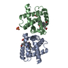
- PDB-6q56: Crystal structure of the B. subtilis M1A22 tRNA methyltransferase TrmK -
+ Open data

- Basic information
| Entry | Database: PDB / ID: 6q56 | ||||||
|---|---|---|---|---|---|---|---|
| Title | Crystal structure of the B. subtilis M1A22 tRNA methyltransferase TrmK | ||||||
 Components Components | tRNA (adenine(22)-N(1))-methyltransferase | ||||||
 Keywords Keywords |  RNA BINDING PROTEIN / methyltransferase / tRNA methyltransferase RNA BINDING PROTEIN / methyltransferase / tRNA methyltransferase | ||||||
| Function / homology |  Function and homology informationtRNA (adenine22-N1)-methyltransferase / : / tRNA methylation / cytoplasm Function and homology informationtRNA (adenine22-N1)-methyltransferase / : / tRNA methylation / cytoplasmSimilarity search - Function | ||||||
| Biological species |  Bacillus subtilis (bacteria) Bacillus subtilis (bacteria) | ||||||
| Method | X-RAY DIFFRACTION / SYNCHROTRON / MOLECULAR REPLACEMENT / Resolution: 2 Å | ||||||
 Authors Authors | Degut, C. / Roovers, M. / Barraud, P. / Brachet, F. / Feller, A. / Larue, V. / Al Refaii, A. / Caillet, J. / Droogmans, L. / Tisne, C. | ||||||
 Citation Citation | Journal: Nucleic Acids Res. / Year: 2019 Title: Structural characterization of B. subtilis m1A22 tRNA methyltransferase TrmK: insights into tRNA recognition. Authors: Degut, C. / Roovers, M. / Barraud, P. / Brachet, F. / Feller, A. / Larue, V. / Al Refaii, A. / Caillet, J. / Droogmans, L. / Tisne, C. #1: Journal: Biomol NMR Assign / Year: 2016Title: Backbone resonance assignments of the m1A22 tRNA methyltransferase TrmK from Bacillus subtilis. Authors: Degut, C. / Barraud, P. / Larue, V. / Tisne, C. | ||||||
| History |
|
- Structure visualization
| Structure viewer | Molecule: MolmilJmol/JSmol |
|---|
- Downloads & links
-Download
| PDBx/mmCIF format | 6q56.cif.gz | 193.4 KB | Display | PDBx/mmCIF format |
|---|---|---|---|---|
| PDB format | pdb6q56.ent.gz | 155.3 KB | Display | PDB format |
| PDBx/mmJSON format | 6q56.json.gz | Tree view | PDBx/mmJSON format | |
| Others |  Other downloads Other downloads |
-Validation report
| Arichive directory | https://data.pdbj.org/pub/pdb/validation_reports/q5/6q56ftp://data.pdbj.org/pub/pdb/validation_reports/q5/6q56 | HTTPS FTP |
|---|
-Related structure data
| Related structure data | 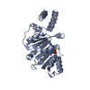 3ku1S S: Starting model for refinement |
|---|---|
| Similar structure data |


-Links
 PDBj
PDBj- Assembly
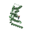
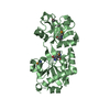
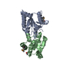
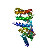
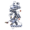
| Deposited unit | 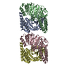
| ||||||||
|---|---|---|---|---|---|---|---|---|---|
| 1 | 
| ||||||||
| 2 | 
| ||||||||
| 3 | 
| ||||||||
| 4 | 
| ||||||||
| Unit cell |
|
-Components
| #1: Protein | Mass: 26165.803 Da / Num. of mol.: 4 / Mutation: C35S C152S Source method: isolated from a genetically manipulated source Source: (gene. exp.) Bacillus subtilis (strain 168) (bacteria)Gene: trmK, yqfN, BSU25180 / Production host: Escherichia coli (E. coli) / Strain (production host): BL21(DE3)References: UniProt: P54471, tRNA (adenine22-N1)-methyltransferase#2: Chemical | ChemComp-NI / Nickel  Mass: 58.693 Da / Num. of mol.: 4 / Source method: obtained synthetically / Formula: Ni Mass: 58.693 Da / Num. of mol.: 4 / Source method: obtained synthetically / Formula: Ni#3: Water | ChemComp-HOH / | Water Mass: 18.015 Da / Num. of mol.: 303 / Source method: isolated from a natural source / Formula: H2O Mass: 18.015 Da / Num. of mol.: 303 / Source method: isolated from a natural source / Formula: H2O |
|---|
-Experimental details
-Experiment
| Experiment | Method: X-RAY DIFFRACTION / Number of used crystals: 1 |
|---|
- Sample preparation
| Crystal | Density Matthews: 2.05 Å3/Da / Density % sol: 40 % |
|---|---|
| Crystal grow | Temperature: 277 K / Method: vapor diffusion, hanging drop / pH: 5 Details: 0.2 M ammonium acetate 0.1 M sodium acetate trihydrate pH 5 30% w/v polyethylene glycol 4000 8% 2-Methyl-2,4-pentanediol |
-Data collection
| Diffraction | Mean temperature: 80 K / Serial crystal experiment: N |
|---|---|
| Diffraction source | Source: SYNCHROTRON / Site: ESRF  / Beamline: ID14-1 / Wavelength: 0.9334 Å / Beamline: ID14-1 / Wavelength: 0.9334 Å |
| Detector | Type: ADSC QUANTUM 210 / Detector: CCD / Date: Sep 30, 2011 |
| Radiation | Protocol: SINGLE WAVELENGTH / Monochromatic (M) / Laue (L): M / Scattering type: x-ray |
| Radiation wavelength | Wavelength: 0.9334 Å / Relative weight: 1 |
| Reflection | Resolution: 1.98→46.24 Å / Num. obs: 57747 / % possible obs: 100 % / Redundancy: 3.8 % / Biso Wilson estimate: 32.71 Å2 / Rmerge(I) obs: 0.2108 / Net I/σ(I): 12 |
| Reflection shell | Resolution: 1.98→2.175 Å / Redundancy: 3.8 % / Rmerge(I) obs: 1.451 / Mean I/σ(I) obs: 1.11 / Num. unique obs: 4968 / CC1/2: 0.227 / % possible all: 100 |
- Processing
| Software |
| ||||||||||||||||||||||||||||||||||||||||||||||||||||||||||||||||||||||||||||||||||||||||||||||||||||||||||||||||||||||||||||||||||||||||||||||||||||||||||
|---|---|---|---|---|---|---|---|---|---|---|---|---|---|---|---|---|---|---|---|---|---|---|---|---|---|---|---|---|---|---|---|---|---|---|---|---|---|---|---|---|---|---|---|---|---|---|---|---|---|---|---|---|---|---|---|---|---|---|---|---|---|---|---|---|---|---|---|---|---|---|---|---|---|---|---|---|---|---|---|---|---|---|---|---|---|---|---|---|---|---|---|---|---|---|---|---|---|---|---|---|---|---|---|---|---|---|---|---|---|---|---|---|---|---|---|---|---|---|---|---|---|---|---|---|---|---|---|---|---|---|---|---|---|---|---|---|---|---|---|---|---|---|---|---|---|---|---|---|---|---|---|---|---|---|---|
| Refinement | Method to determine structure: MOLECULAR REPLACEMENT Starting model: 3KU1 Resolution: 2→46.239 Å / SU ML: 0.33 / Cross valid method: FREE R-VALUE / σ(F): 1.34 / Phase error: 27.87 / Stereochemistry target values: ML
| ||||||||||||||||||||||||||||||||||||||||||||||||||||||||||||||||||||||||||||||||||||||||||||||||||||||||||||||||||||||||||||||||||||||||||||||||||||||||||
| Solvent computation | Shrinkage radii: 0.9 Å / VDW probe radii: 1.11 Å / Solvent model: FLAT BULK SOLVENT MODEL | ||||||||||||||||||||||||||||||||||||||||||||||||||||||||||||||||||||||||||||||||||||||||||||||||||||||||||||||||||||||||||||||||||||||||||||||||||||||||||
| Refinement step | Cycle: LAST / Resolution: 2→46.239 Å
| ||||||||||||||||||||||||||||||||||||||||||||||||||||||||||||||||||||||||||||||||||||||||||||||||||||||||||||||||||||||||||||||||||||||||||||||||||||||||||
| Refine LS restraints |
| ||||||||||||||||||||||||||||||||||||||||||||||||||||||||||||||||||||||||||||||||||||||||||||||||||||||||||||||||||||||||||||||||||||||||||||||||||||||||||
| LS refinement shell |
|
