 Movie
Movie Controller

[English] 日本語
 Yorodumi
- PDB-6ovh: Cryo-EM structure of Bimetallic dodecameric cage design 3 (BMC3) ... -
+ Open data

- Basic information
| Entry | Database: PDB / ID: 6ovh | |||||||||||||||
|---|---|---|---|---|---|---|---|---|---|---|---|---|---|---|---|---|
| Title | Cryo-EM structure of Bimetallic dodecameric cage design 3 (BMC3) from cytochrome cb562 | |||||||||||||||
 Components Components | Soluble cytochrome b562 | |||||||||||||||
 Keywords Keywords | METAL BINDING PROTEIN /  Supramolecular assembly / protein cage / bimetallic / metal binding / hydroxamic acid Supramolecular assembly / protein cage / bimetallic / metal binding / hydroxamic acid | |||||||||||||||
| Function / homology |  Function and homology informationelectron transfer activity / periplasmic space / iron ion binding / heme binding Function and homology informationelectron transfer activity / periplasmic space / iron ion binding / heme bindingSimilarity search - Function | |||||||||||||||
| Biological species |  Escherichia coli (E. coli) Escherichia coli (E. coli) | |||||||||||||||
| Method | ELECTRON MICROSCOPY / single particle reconstruction / cryo EM / Resolution: 2.6 Å | |||||||||||||||
| Model details | Keywords: Supramolecular assembly, protein cage, bimetallic, metal binding | |||||||||||||||
 Authors Authors | Golub, E. / Subramanian, R.H. / Yan, X. / Alberstein, R.G. / Tezcan, F.A. | |||||||||||||||
| Funding support |  United States, European Union, United States, European Union,  Germany, 4items Germany, 4items
| |||||||||||||||
 Citation Citation | Journal: Nature / Year: 2020 Title: Constructing protein polyhedra via orthogonal chemical interactions. Authors: Eyal Golub / Rohit H Subramanian / Julian Esselborn / Robert G Alberstein / Jake B Bailey / Jerika A Chiong / Xiaodong Yan / Timothy Booth / Timothy S Baker / F Akif Tezcan / Abstract: Many proteins exist naturally as symmetrical homooligomers or homopolymers. The emergent structural and functional properties of such protein assemblies have inspired extensive efforts in ...Many proteins exist naturally as symmetrical homooligomers or homopolymers. The emergent structural and functional properties of such protein assemblies have inspired extensive efforts in biomolecular design. As synthesized by ribosomes, proteins are inherently asymmetric. Thus, they must acquire multiple surface patches that selectively associate to generate the different symmetry elements needed to form higher-order architectures-a daunting task for protein design. Here we address this problem using an inorganic chemical approach, whereby multiple modes of protein-protein interactions and symmetry are simultaneously achieved by selective, 'one-pot' coordination of soft and hard metal ions. We show that a monomeric protein (protomer) appropriately modified with biologically inspired hydroxamate groups and zinc-binding motifs assembles through concurrent Fe and Zn coordination into discrete dodecameric and hexameric cages. Our cages closely resemble natural polyhedral protein architectures and are, to our knowledge, unique among designed systems in that they possess tightly packed shells devoid of large apertures. At the same time, they can assemble and disassemble in response to diverse stimuli, owing to their heterobimetallic construction on minimal interprotein-bonding footprints. With stoichiometries ranging from [2 Fe:9 Zn:6 protomers] to [8 Fe:21 Zn:12 protomers], these protein cages represent some of the compositionally most complex protein assemblies-or inorganic coordination complexes-obtained by design. | |||||||||||||||
| History |
|
- Structure visualization
| Movie |
Movie viewer |
|---|---|
| Structure viewer | Molecule: MolmilJmol/JSmol |
- Downloads & links
-Download
| PDBx/mmCIF format | 6ovh.cif.gz | 256.1 KB | Display | PDBx/mmCIF format |
|---|---|---|---|---|
| PDB format | pdb6ovh.ent.gz | 217.3 KB | Display | PDB format |
| PDBx/mmJSON format | 6ovh.json.gz | Tree view | PDBx/mmJSON format | |
| Others |  Other downloads Other downloads |
-Validation report
| Arichive directory | https://data.pdbj.org/pub/pdb/validation_reports/ov/6ovhftp://data.pdbj.org/pub/pdb/validation_reports/ov/6ovh | HTTPS FTP |
|---|
-Related structure data
| Related structure data |  20212MC  6ot4C 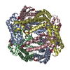 6ot7C  6ot8C  6ot9C M: map data used to model this data C: citing same article ( |
|---|---|
| Similar structure data |


-Links
 PDBj
PDBj


- Assembly
| Deposited unit | 
|
|---|---|
| 1 |
|
-Components
-Protein , 1 types, 12 molecules ABCDEFGHIJKL
| #1: Protein | Mass: 11809.307 Da / Num. of mol.: 12 Mutation: D5H,E8H,V16H,A24T,Q25T,R34Q,L38Q,Q41W,K42S,K59S,H63C,D66W,I67E,V69I,D73N,D74A,K77H,N80K,E81Q,G82C,R98C,Y101C Source method: isolated from a genetically manipulated source Details: pET20b for expression of BMC3 described here with background of pEC86 to provide machinery for c-type linkage of heme. Source: (gene. exp.) Escherichia coli (E. coli) / Gene: cybC / Plasmid: pET20b-BMC3/pEC86Details (production host): pET20b for expression of BMC3 described here with background of pEC86 to provide machinery for c-type linkage of heme. Production host: Escherichia coli BL21(DE3) (bacteria) / Variant (production host): BL21(DE3) / References: UniProt: P0ABE7 |
|---|
-Non-polymers , 5 types, 239 molecules 


| #2: Chemical | ChemComp-HEC / Heme C Mass: 618.503 Da / Num. of mol.: 12 / Source method: obtained synthetically / Formula: C34H34FeN4O4 Mass: 618.503 Da / Num. of mol.: 12 / Source method: obtained synthetically / Formula: C34H34FeN4O4#3: Chemical | ChemComp-HAE / Acetohydroxamic acid Mass: 75.067 Da / Num. of mol.: 24 / Source method: obtained synthetically / Formula: C2H5NO2 / Comment: inhibitor, medication*YM Mass: 75.067 Da / Num. of mol.: 24 / Source method: obtained synthetically / Formula: C2H5NO2 / Comment: inhibitor, medication*YM#4: Chemical | ChemComp-ZN /  Mass: 65.409 Da / Num. of mol.: 24 / Source method: obtained synthetically / Formula: Zn Mass: 65.409 Da / Num. of mol.: 24 / Source method: obtained synthetically / Formula: Zn#5: Chemical | ChemComp-FE / Iron Mass: 55.845 Da / Num. of mol.: 8 / Source method: obtained synthetically / Formula: Fe Mass: 55.845 Da / Num. of mol.: 8 / Source method: obtained synthetically / Formula: Fe#6: Water | ChemComp-HOH / | WaterMass: 18.015 Da / Num. of mol.: 171 / Source method: isolated from a natural source / Formula: H2O |
|---|
-Experimental details
-Experiment
| Experiment | Method: ELECTRON MICROSCOPY |
|---|---|
| EM experiment | Aggregation state: PARTICLE / 3D reconstruction method: single particle reconstruction |
- Sample preparation
| Component | Name: Bimetallic dodecameric cage 3 (BMC3) / Type: COMPLEX Details: Cryo-EM reconstruction of self-assembled BMC3 dodecameric cages Entity ID: #1 / Source: RECOMBINANT |
|---|---|
| Molecular weight | Value: 0.15 MDa / Experimental value: YES |
| Source (natural) | Organism: Escherichia coli (E. coli) |
| Source (recombinant) | Organism: Escherichia coli BL21(DE3) (bacteria) / Plasmid: pET20b-BMC3/pEC86 |
| Buffer solution | pH: 8.5 |
| Buffer component | Conc.: 20 mM / Name: Tris |
| Specimen | Embedding applied: NO / Shadowing applied: NO / Staining applied: NO / Vitrification applied: YES Details: Protein solutions containing 20 micromolar BMC3 in 20 mM Tris (pH 8.5) were incubated with [FeSO4] = 20 micromolar, [ZnCl2] = 60 micromolar for 2-3 h to form cages. Samples were concentrated ...Details: Protein solutions containing 20 micromolar BMC3 in 20 mM Tris (pH 8.5) were incubated with [FeSO4] = 20 micromolar, [ZnCl2] = 60 micromolar for 2-3 h to form cages. Samples were concentrated 10 fold prior to grid preparation. |
| Specimen support | Details: The grid was glow discharged at 20 mA for 30 s. / Grid material: COPPER / Grid mesh size: 400 divisions/in. / Grid type: Quantifoil R1.2/1.3 |
| Vitrification | Instrument: FEI VITROBOT MARK IV / Cryogen name: ETHANE |
- Electron microscopy imaging
| Experimental equipment |  Model: Titan Krios / Image courtesy: FEI Company |
|---|---|
| Microscopy | Model: FEI TITAN KRIOS |
| Electron gun | Electron source: FIELD EMISSION GUN / Accelerating voltage: 300 kV / Illumination mode: FLOOD BEAM |
| Electron lens | Mode: BRIGHT FIELDBright-field microscopy |
| Image recording | Average exposure time: 10 sec. / Electron dose: 60 e/Å2 / Film or detector model: GATAN K2 SUMMIT (4k x 4k) / Num. of real images: 4672 |
| Image scans | Movie frames/image: 50 |
- Processing
| Software |
| ||||||||||||||||||||||||
|---|---|---|---|---|---|---|---|---|---|---|---|---|---|---|---|---|---|---|---|---|---|---|---|---|---|
| EM software |
| ||||||||||||||||||||||||
| CTF correction | Type: NONE | ||||||||||||||||||||||||
| Particle selection | Num. of particles selected: 805156 | ||||||||||||||||||||||||
| Symmetry | Point symmetry: T (tetrahedral) | ||||||||||||||||||||||||
| 3D reconstruction | Resolution: 2.6 Å / Resolution method: FSC 0.143 CUT-OFF / Num. of particles: 25391 / Symmetry type: POINT | ||||||||||||||||||||||||
| Atomic model building | Protocol: RIGID BODY FIT / Space: REAL Details: Symmetry mates were generated from the atomic model to build the dodecameric cage. Chain IDs were re-assigned to ascend from A-L. The cage PDB was manually fit to the EM density map in UCSF ...Details: Symmetry mates were generated from the atomic model to build the dodecameric cage. Chain IDs were re-assigned to ascend from A-L. The cage PDB was manually fit to the EM density map in UCSF Chimera and refined using phenix.real_space_refine | ||||||||||||||||||||||||
| Atomic model building | PDB-ID: 6OT7 | ||||||||||||||||||||||||
| Refinement | Stereochemistry target values: CDL v1.2 | ||||||||||||||||||||||||
| Refine LS restraints |
|
