National Institutes of Health/National Institute of General Medical Sciences (NIH/NIGMS)
R35GM122510
United States
Citation
Journal: Proc Natl Acad Sci U S A / Year: 2019 Title: A packing for A-form DNA in an icosahedral virus. Authors: Fengbin Wang / Ying Liu / Zhangli Su / Tomasz Osinski / Guilherme A P de Oliveira / James F Conway / Stefan Schouten / Mart Krupovic / David Prangishvili / Edward H Egelman / Abstract: Studies on viruses infecting archaea living in the most extreme environments continue to show a remarkable diversity of structures, suggesting that the sampling continues to be very sparse. We have ...Studies on viruses infecting archaea living in the most extreme environments continue to show a remarkable diversity of structures, suggesting that the sampling continues to be very sparse. We have used electron cryo-microscopy to study at 3.7-Å resolution the structure of the polyhedral virus 1 (SPV1), which was originally isolated from a hot, acidic spring in Beppu, Japan. The 2 capsid proteins with variant single jelly-roll folds form pentamers and hexamers which assemble into a = 43 icosahedral shell. In contrast to tailed icosahedral double-stranded DNA (dsDNA) viruses infecting bacteria and archaea, and herpesviruses infecting animals and humans, where naked DNA is packed under very high pressure due to the repulsion between adjacent layers of DNA, the circular dsDNA in SPV1 is fully covered with a viral protein forming a nucleoprotein filament with attractive interactions between layers. Most strikingly, we have been able to show that the DNA is in an A-form, as it is in the filamentous viruses infecting hyperthermophilic acidophiles. Previous studies have suggested that DNA is in the B-form in bacteriophages, and our study is a direct visualization of the structure of DNA in an icosahedral virus.
T: Structural protein VP4 U: Structural protein VP4 V: Structural protein VP4 W: Structural protein VP4 X: Structural protein VP4 G: Structural protein VP4 n: Structural protein VP4 o: Structural protein VP4 p: Structural protein VP4 q: Structural protein VP4 r: Structural protein VP4 g: Structural protein VP4 N: Structural protein VP4 O: Structural protein VP4 P: Structural protein VP4 Q: Structural protein VP4 R: Structural protein VP4 S: Structural protein VP4 h: Structural protein VP4 i: Structural protein VP4 j: Structural protein VP4 k: Structural protein VP4 l: Structural protein VP4 m: Structural protein VP4 H: Structural protein VP4 I: Structural protein VP4 J: Structural protein VP4 K: Structural protein VP4 L: Structural protein VP4 M: Structural protein VP4 a: Structural protein VP4 b: Structural protein VP4 c: Structural protein VP4 d: Structural protein VP4 e: Structural protein VP4 f: Structural protein VP4 A: Structural protein VP4 B: Structural protein VP4 C: Structural protein VP4 D: Structural protein VP4 E: Structural protein VP4 F: Structural protein VP4 Z: Uncharacterized protein
T: Structural protein VP4 U: Structural protein VP4 V: Structural protein VP4 W: Structural protein VP4 X: Structural protein VP4 G: Structural protein VP4 n: Structural protein VP4 o: Structural protein VP4 p: Structural protein VP4 q: Structural protein VP4 r: Structural protein VP4 g: Structural protein VP4 N: Structural protein VP4 O: Structural protein VP4 P: Structural protein VP4 Q: Structural protein VP4 R: Structural protein VP4 S: Structural protein VP4 h: Structural protein VP4 i: Structural protein VP4 j: Structural protein VP4 k: Structural protein VP4 l: Structural protein VP4 m: Structural protein VP4 H: Structural protein VP4 I: Structural protein VP4 J: Structural protein VP4 K: Structural protein VP4 L: Structural protein VP4 M: Structural protein VP4 a: Structural protein VP4 b: Structural protein VP4 c: Structural protein VP4 d: Structural protein VP4 e: Structural protein VP4 f: Structural protein VP4 A: Structural protein VP4 B: Structural protein VP4 C: Structural protein VP4 D: Structural protein VP4 E: Structural protein VP4 F: Structural protein VP4 Z: Uncharacterized protein
Idetical with deposited unit in distinct coordinate
icosahedral asymmetric unit
Type
Name
Symmetry operation
Number
point symmetry operation
1
3
T: Structural protein VP4 U: Structural protein VP4 V: Structural protein VP4 W: Structural protein VP4 X: Structural protein VP4 G: Structural protein VP4 n: Structural protein VP4 o: Structural protein VP4 p: Structural protein VP4 q: Structural protein VP4 r: Structural protein VP4 g: Structural protein VP4 N: Structural protein VP4 O: Structural protein VP4 P: Structural protein VP4 Q: Structural protein VP4 R: Structural protein VP4 S: Structural protein VP4 h: Structural protein VP4 i: Structural protein VP4 j: Structural protein VP4 k: Structural protein VP4 l: Structural protein VP4 m: Structural protein VP4 H: Structural protein VP4 I: Structural protein VP4 J: Structural protein VP4 K: Structural protein VP4 L: Structural protein VP4 M: Structural protein VP4 a: Structural protein VP4 b: Structural protein VP4 c: Structural protein VP4 d: Structural protein VP4 e: Structural protein VP4 f: Structural protein VP4 A: Structural protein VP4 B: Structural protein VP4 C: Structural protein VP4 D: Structural protein VP4 E: Structural protein VP4 F: Structural protein VP4 Z: Uncharacterized protein
x 5
icosahedral pentamer
4.18 MDa, 215 polymers
Theoretical mass
Number of molelcules
Total (without water)
4,176,012
215
Polymers
4,176,012
215
Non-polymers
0
0
Water
0
Type
Name
Symmetry operation
Number
point symmetry operation
5
4
T: Structural protein VP4 U: Structural protein VP4 V: Structural protein VP4 W: Structural protein VP4 X: Structural protein VP4 G: Structural protein VP4 n: Structural protein VP4 o: Structural protein VP4 p: Structural protein VP4 q: Structural protein VP4 r: Structural protein VP4 g: Structural protein VP4 N: Structural protein VP4 O: Structural protein VP4 P: Structural protein VP4 Q: Structural protein VP4 R: Structural protein VP4 S: Structural protein VP4 h: Structural protein VP4 i: Structural protein VP4 j: Structural protein VP4 k: Structural protein VP4 l: Structural protein VP4 m: Structural protein VP4 H: Structural protein VP4 I: Structural protein VP4 J: Structural protein VP4 K: Structural protein VP4 L: Structural protein VP4 M: Structural protein VP4 a: Structural protein VP4 b: Structural protein VP4 c: Structural protein VP4 d: Structural protein VP4 e: Structural protein VP4 f: Structural protein VP4 A: Structural protein VP4 B: Structural protein VP4 C: Structural protein VP4 D: Structural protein VP4 E: Structural protein VP4 F: Structural protein VP4 Z: Uncharacterized protein
x 6
icosahedral 23 hexamer
5.01 MDa, 258 polymers
Theoretical mass
Number of molelcules
Total (without water)
5,011,215
258
Polymers
5,011,215
258
Non-polymers
0
0
Water
0
Type
Name
Symmetry operation
Number
point symmetry operation
6
5
Idetical with deposited unit in distinct coordinate
icosahedral asymmetric unit, std point frame
Type
Name
Symmetry operation
Number
transform to point frame
1
Symmetry
Point symmetry: (Schoenflies symbol: I (icosahedral))
-
Components
#1: Protein
... StructuralproteinVP4 / Structure
Mass: 19394.885 Da / Num. of mol.: 42 / Source method: isolated from a natural source / Source: (natural) Sulfolobus polyhedral virus 1 / References: UniProt: A0A1W6I187
#2: Protein
Uncharacterizedprotein
Mass: 20617.277 Da / Num. of mol.: 1 / Source method: isolated from a natural source / Source: (natural) Sulfolobus polyhedral virus 1 / References: UniProt: A0A1W6I162
-
Experimental details
-
Experiment
Experiment
Method: ELECTRON MICROSCOPY
EM experiment
Aggregation state: PARTICLE / 3D reconstruction method: single particle reconstruction
In the structure databanks used in Yorodumi, some data are registered as the other names, "COVID-19 virus" and "2019-nCoV". Here are the details of the virus and the list of structure data.
Jan 31, 2019. EMDB accession codes are about to change! (news from PDBe EMDB page)
EMDB accession codes are about to change! (news from PDBe EMDB page)
The allocation of 4 digits for EMDB accession codes will soon come to an end. Whilst these codes will remain in use, new EMDB accession codes will include an additional digit and will expand incrementally as the available range of codes is exhausted. The current 4-digit format prefixed with “EMD-” (i.e. EMD-XXXX) will advance to a 5-digit format (i.e. EMD-XXXXX), and so on. It is currently estimated that the 4-digit codes will be depleted around Spring 2019, at which point the 5-digit format will come into force.
The EM Navigator/Yorodumi systems omit the EMD- prefix.
Related info.:Q: What is EMD? / ID/Accession-code notation in Yorodumi/EM Navigator
Yorodumi is a browser for structure data from EMDB, PDB, SASBDB, etc.
This page is also the successor to EM Navigator detail page, and also detail information page/front-end page for Omokage search.
The word "yorodu" (or yorozu) is an old Japanese word meaning "ten thousand". "mi" (miru) is to see.
Related info.:EMDB / PDB / SASBDB / Comparison of 3 databanks / Yorodumi Search / Aug 31, 2016. New EM Navigator & Yorodumi / Yorodumi Papers / Jmol/JSmol / Function and homology information / Changes in new EM Navigator and Yorodumi
 Movie
Movie Controller


 Open data
 Basic information
 Components
 Keywords
 VIRUS / archaeal pilus /
  Function and homology information
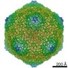
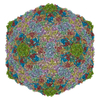
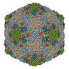
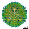
 Sulfolobus polyhedral virus 1
 Authors
 United States, 1items
  Citation

 Structure visualization
 Downloads & links
 Other downloads


 PDBj
PDBj Assembly


 Sample preparation
 Electron microscopy imaging
 Processing