 Movie
Movie Controller

[English] 日本語
 Yorodumi
- PDB-6nob: Structure of Glycoside Hydrolase family 32 from Bifidobacterium a... -
+ Open data

- Basic information
| Entry | Database: PDB / ID: 6nob | ||||||||||||
|---|---|---|---|---|---|---|---|---|---|---|---|---|---|
| Title | Structure of Glycoside Hydrolase family 32 from Bifidobacterium adolescentis | ||||||||||||
 Components Components | Beta-fructofuranosidase Sucrase Sucrase | ||||||||||||
 Keywords Keywords | HYDROLASE / GH32 / glycoside hydrolase | ||||||||||||
| Function / homology |  Function and homology information Function and homology informationhydrolase activity, hydrolyzing O-glycosyl compounds / carbohydrate metabolic process Similarity search - Function | ||||||||||||
| Biological species |  Bifidobacterium adolescentis (bacteria) Bifidobacterium adolescentis (bacteria) | ||||||||||||
| Method | X-RAY DIFFRACTION / SYNCHROTRON / MOLECULAR REPLACEMENT / Resolution: 2.44 Å | ||||||||||||
 Authors Authors | Mera, A.M. / Lima, M.Z.T. / Muniz, J.R.C. | ||||||||||||
| Funding support |  Brazil, 3items Brazil, 3items
| ||||||||||||
 Citation Citation | Journal: To Be Published Title: Structure of GH32 from Bifidobacterium adolescentis Authors: Mera, A. / Lima, M.Z.T. / Bernardes, A. / Garcia, W. / Muniz, J.R.C. | ||||||||||||
| History |
|
- Structure visualization
| Structure viewer | Molecule: MolmilJmol/JSmol |
|---|
- Downloads & links
-Download
| PDBx/mmCIF format | 6nob.cif.gz | 317.1 KB | Display | PDBx/mmCIF format |
|---|---|---|---|---|
| PDB format | pdb6nob.ent.gz | 215.1 KB | Display | PDB format |
| PDBx/mmJSON format | 6nob.json.gz | Tree view | PDBx/mmJSON format | |
| Others |  Other downloads Other downloads |
-Validation report
| Arichive directory | https://data.pdbj.org/pub/pdb/validation_reports/no/6nobftp://data.pdbj.org/pub/pdb/validation_reports/no/6nob | HTTPS FTP |
|---|
-Related structure data
| Related structure data |  1uypS S: Starting model for refinement |
|---|---|
| Similar structure data |


-Links
 PDBj
PDBj- Assembly
| Deposited unit | 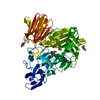
| ||||||||||||
|---|---|---|---|---|---|---|---|---|---|---|---|---|---|
| 1 |
| ||||||||||||
| 2 | 
| ||||||||||||
| Unit cell |
| ||||||||||||
| Components on special symmetry positions |
|
-Components
| #1: Protein | Sucrase Mass: 70221.898 Da / Num. of mol.: 1 Source method: isolated from a genetically manipulated source Source: (gene. exp.) Bifidobacterium adolescentis (strain ATCC 15703 / DSM 20083 / NCTC 11814 / E194a) (bacteria)Strain: ATCC 15703 / DSM 20083 / NCTC 11814 / E194a / Gene: BAD_1325 / Production host: Escherichia coli (E. coli) / References: UniProt: A1A323 | ||||
|---|---|---|---|---|---|
| #2: Chemical | ChemComp-EDO / Ethylene glycol  Mass: 62.068 Da / Num. of mol.: 4 / Source method: obtained synthetically / Formula: C2H6O2 Mass: 62.068 Da / Num. of mol.: 4 / Source method: obtained synthetically / Formula: C2H6O2#3: Chemical | Diethylene glycol  Mass: 106.120 Da / Num. of mol.: 3 / Source method: obtained synthetically / Formula: C4H10O3 Mass: 106.120 Da / Num. of mol.: 3 / Source method: obtained synthetically / Formula: C4H10O3#4: Water | ChemComp-HOH / | Water Mass: 18.015 Da / Num. of mol.: 374 / Source method: isolated from a natural source / Formula: H2O Mass: 18.015 Da / Num. of mol.: 374 / Source method: isolated from a natural source / Formula: H2O |
-Experimental details
-Experiment
| Experiment | Method: X-RAY DIFFRACTION / Number of used crystals: 1 |
|---|
- Sample preparation
| Crystal | Density Matthews: 4.7 Å3/Da / Density % sol: 73.85 % / Description: hexagonal crystals |
|---|---|
| Crystal grow | Temperature: 291.15 K / Method: vapor diffusion, sitting drop / pH: 6.5 / Details: PEG 8K 25% (w/v), MES 0.1M pH 6.5 |
-Data collection
| Diffraction | Mean temperature: 100 K / Serial crystal experiment: N |
|---|---|
| Diffraction source | Source: SYNCHROTRON / Site: LNLS / Beamline: W01B-MX2 / Wavelength: 1.45866 Å |
| Detector | Type: DECTRIS PILATUS 2M / Detector: PIXEL / Date: Nov 6, 2015 |
| Radiation | Protocol: SINGLE WAVELENGTH / Monochromatic (M) / Laue (L): M / Scattering type: x-ray |
| Radiation wavelength | Wavelength: 1.45866 Å / Relative weight: 1 |
| Reflection | Resolution: 2.44→57.34 Å / Num. obs: 49401 / % possible obs: 100 % / Redundancy: 18.4 % / Biso Wilson estimate: 42.84 Å2 / CC1/2: 0.996 / Rmerge(I) obs: 0.282 / Rrim(I) all: 0.298 / Net I/σ(I): 9.9 |
| Reflection shell | Resolution: 2.44→2.5 Å / Redundancy: 15.1 % / Rmerge(I) obs: 3.425 / Mean I/σ(I) obs: 1 / Num. unique obs: 54042 / CC1/2: 0.669 / Rpim(I) all: 0.926 / Rrim(I) all: 3.67 / % possible all: 100 |
- Processing
| Software |
| |||||||||||||||||||||||||||||||||||||||||||||||||||||||||||||||||||||||||||||||||||||||||||||||||||||||||||||||||||||||||||||||||||||
|---|---|---|---|---|---|---|---|---|---|---|---|---|---|---|---|---|---|---|---|---|---|---|---|---|---|---|---|---|---|---|---|---|---|---|---|---|---|---|---|---|---|---|---|---|---|---|---|---|---|---|---|---|---|---|---|---|---|---|---|---|---|---|---|---|---|---|---|---|---|---|---|---|---|---|---|---|---|---|---|---|---|---|---|---|---|---|---|---|---|---|---|---|---|---|---|---|---|---|---|---|---|---|---|---|---|---|---|---|---|---|---|---|---|---|---|---|---|---|---|---|---|---|---|---|---|---|---|---|---|---|---|---|---|---|
| Refinement | Method to determine structure: MOLECULAR REPLACEMENT Starting model: 1UYP Resolution: 2.44→52.14 Å / SU ML: 0.2749 / Cross valid method: FREE R-VALUE / σ(F): 1.33 / Phase error: 23.4693
| |||||||||||||||||||||||||||||||||||||||||||||||||||||||||||||||||||||||||||||||||||||||||||||||||||||||||||||||||||||||||||||||||||||
| Solvent computation | Shrinkage radii: 0.9 Å / VDW probe radii: 1.11 Å | |||||||||||||||||||||||||||||||||||||||||||||||||||||||||||||||||||||||||||||||||||||||||||||||||||||||||||||||||||||||||||||||||||||
| Displacement parameters | Biso mean: 46.94 Å2 | |||||||||||||||||||||||||||||||||||||||||||||||||||||||||||||||||||||||||||||||||||||||||||||||||||||||||||||||||||||||||||||||||||||
| Refinement step | Cycle: LAST / Resolution: 2.44→52.14 Å
| |||||||||||||||||||||||||||||||||||||||||||||||||||||||||||||||||||||||||||||||||||||||||||||||||||||||||||||||||||||||||||||||||||||
| Refine LS restraints |
| |||||||||||||||||||||||||||||||||||||||||||||||||||||||||||||||||||||||||||||||||||||||||||||||||||||||||||||||||||||||||||||||||||||
| LS refinement shell |
| |||||||||||||||||||||||||||||||||||||||||||||||||||||||||||||||||||||||||||||||||||||||||||||||||||||||||||||||||||||||||||||||||||||
| Refinement TLS params. | Method: refined / Refine-ID: X-RAY DIFFRACTION
| |||||||||||||||||||||||||||||||||||||||||||||||||||||||||||||||||||||||||||||||||||||||||||||||||||||||||||||||||||||||||||||||||||||
| Refinement TLS group |
|
