 Movie
Movie Controller

[English] 日本語
 Yorodumi
- PDB-6myt: Avian mitochondrial complex II with Atpenin A5 bound, sidechain i... -
+ Open data

- Basic information
| Entry | Database: PDB / ID: 6myt | ||||||
|---|---|---|---|---|---|---|---|
| Title | Avian mitochondrial complex II with Atpenin A5 bound, sidechain in pocket | ||||||
 Components Components |
| ||||||
 Keywords Keywords | OXIDOREDUCTASE/OXIDOREDUCTASE INHIBITOR /  Complex II / membrane protein / heme protein / OXIDOREDUCTASE-OXIDOREDUCTASE INHIBITOR complex Complex II / membrane protein / heme protein / OXIDOREDUCTASE-OXIDOREDUCTASE INHIBITOR complex | ||||||
| Function / homology |  Function and homology information Function and homology informationCitric acid cycle (TCA cycle) / The tricarboxylic acid cycle / succinate dehydrogenase complex / succinate dehydrogenase activity / succinate metabolic process / mitochondrial electron transport, succinate to ubiquinone / mitochondrial respiratory chain complex II, succinate dehydrogenase complex (ubiquinone) / succinate dehydrogenase (quinone) activity / : / succinate dehydrogenase ...Citric acid cycle (TCA cycle) / The tricarboxylic acid cycle / succinate dehydrogenase complex / succinate dehydrogenase activity / succinate metabolic process / mitochondrial electron transport, succinate to ubiquinone / mitochondrial respiratory chain complex II, succinate dehydrogenase complex (ubiquinone) / succinate dehydrogenase (quinone) activity / : / succinate dehydrogenase / oxidoreductase activity, acting on the CH-CH group of donors / ubiquinone binding / 3 iron, 4 sulfur cluster binding / aerobic respiration / tricarboxylic acid cycle / respiratory electron transport chain / mitochondrial membrane / 2 iron, 2 sulfur cluster binding / flavin adenine dinucleotide binding / 4 iron, 4 sulfur cluster binding / mitochondrial inner membrane / electron transfer activity / heme binding / metal ion bindingSimilarity search - Function | ||||||
| Biological species |  Gallus gallus (chicken) Gallus gallus (chicken) | ||||||
| Method | X-RAY DIFFRACTION / SYNCHROTRON / MOLECULAR REPLACEMENT / Resolution: 2.27 Å | ||||||
 Authors Authors | Berry, E.A. / Huang, L.-S. | ||||||
| Funding support |  United States, 1items United States, 1items
| ||||||
 Citation Citation | Journal: Biochim Biophys Acta Proteins Proteom / Year: 2021 Title: Crystallographic investigation of the ubiquinone binding site of respiratory Complex II and its inhibitors. Authors: Huang, L.S. / Lummen, P. / Berry, E.A. #1: Journal: J. Biol. Chem. / Year: 2006Title: 3-nitropropionic acid is a suicide inhibitor of mitochondrial respiration that, upon oxidation by complex II, forms a covalent adduct with a catalytic base arginine in the active site of the enzyme. Authors: Huang, L.S. / Sun, G. / Cobessi, D. / Wang, A.C. / Shen, J.T. / Tung, E.Y. / Anderson, V.E. / Berry, E.A. #2: Journal: Biochim. Biophys. Acta / Year: 2006Title: Crystallographic studies of the binding of ligands to the dicarboxylate site of Complex II, and the identity of the ligand in the "oxaloacetate-inhibited" state. Authors: Huang, L.S. / Shen, J.T. / Wang, A.C. / Berry, E.A. #3: Journal: Acta Crystallogr. D Biol. Crystallogr. / Year: 2005 Title: Crystallization of mitochondrial respiratory complex II from chicken heart: a membrane-protein complex diffracting to 2.0 A. Authors: Huang, L.S. / Borders, T.M. / Shen, J.T. / Wang, C.J. / Berry, E.A. #4: Journal: Cell / Year: 2005Title: Crystal structure of mitochondrial respiratory membrane protein complex II. Authors: Sun, F. / Huo, X. / Zhai, Y. / Wang, A. / Xu, J. / Su, D. / Bartlam, M. / Rao, Z. | ||||||
| History |
|
- Structure visualization
| Structure viewer | Molecule: MolmilJmol/JSmol |
|---|
- Downloads & links
-Download
| PDBx/mmCIF format | 6myt.cif.gz | 480 KB | Display | PDBx/mmCIF format |
|---|---|---|---|---|
| PDB format | pdb6myt.ent.gz | 386.2 KB | Display | PDB format |
| PDBx/mmJSON format | 6myt.json.gz | Tree view | PDBx/mmJSON format | |
| Others |  Other downloads Other downloads |
-Validation report
| Arichive directory | https://data.pdbj.org/pub/pdb/validation_reports/my/6mytftp://data.pdbj.org/pub/pdb/validation_reports/my/6myt | HTTPS FTP |
|---|
-Related structure data
| Related structure data | 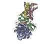 6myoC  6mypC  6myqC  6myrC  6mysC  6myuC  1yq3S S: Starting model for refinement C: citing same article ( |
|---|---|
| Similar structure data |


-Links
 PDBj
PDBj


- Assembly
| Deposited unit | 
| ||||||||
|---|---|---|---|---|---|---|---|---|---|
| 1 |
| ||||||||
| Unit cell |
|
-Components
-Succinate dehydrogenase [ubiquinone] ... , 3 types, 3 molecules ABD
| #1: Protein | Mass: 68256.922 Da / Num. of mol.: 1 / Source method: isolated from a natural source / Source: (natural) Gallus gallus (chicken) / Organ: heartReferences: UniProt: F1NPJ4, UniProt: Q9YHT1*PLUS, succinate dehydrogenase |
|---|---|
| #2: Protein | Mass: 28685.221 Da / Num. of mol.: 1 / Source method: isolated from a natural source / Source: (natural) Gallus gallus (chicken) / Organ: heart / References: UniProt: Q9YHT2, succinate dehydrogenase |
| #4: Protein | Mass: 10971.604 Da / Num. of mol.: 1 / Source method: isolated from a natural source / Source: (natural) Gallus gallus (chicken) / Organ: heart / References: UniProt: Q5ZIS0 |
-Protein , 1 types, 1 molecules C
| #3: Protein | Mass: 15391.153 Da / Num. of mol.: 1 / Source method: isolated from a natural source / Source: (natural) Gallus gallus (chicken) / Organ: heartReferences: UniProt: D0VWW3, UniProt: A0A3Q2U2Y6*PLUS, succinate dehydrogenase |
|---|
-Non-polymers , 13 types, 270 molecules 


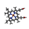


| #5: Chemical | ChemComp-FAD / Flavin adenine dinucleotide Mass: 785.550 Da / Num. of mol.: 1 / Source method: obtained synthetically / Formula: C27H33N9O15P2 / Comment: FAD*YM Mass: 785.550 Da / Num. of mol.: 1 / Source method: obtained synthetically / Formula: C27H33N9O15P2 / Comment: FAD*YM | ||||||||||||||||||
|---|---|---|---|---|---|---|---|---|---|---|---|---|---|---|---|---|---|---|---|
| #6: Chemical | ChemComp-OAA / Oxaloacetic acid Mass: 131.064 Da / Num. of mol.: 1 / Source method: obtained synthetically / Formula: C4H3O5 Mass: 131.064 Da / Num. of mol.: 1 / Source method: obtained synthetically / Formula: C4H3O5 | ||||||||||||||||||
| #7: Chemical | ChemComp-K /  Mass: 39.098 Da / Num. of mol.: 1 / Source method: obtained synthetically / Formula: K Mass: 39.098 Da / Num. of mol.: 1 / Source method: obtained synthetically / Formula: K | ||||||||||||||||||
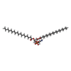
| #8: Chemical | ChemComp-UNL / Num. of mol.: 59 / Source method: obtained synthetically #9: Chemical | ChemComp-PEG / | Diethylene glycol Mass: 106.120 Da / Num. of mol.: 1 / Source method: obtained synthetically / Formula: C4H10O3 Mass: 106.120 Da / Num. of mol.: 1 / Source method: obtained synthetically / Formula: C4H10O3#10: Chemical | ChemComp-FES / | Iron–sulfur cluster Mass: 175.820 Da / Num. of mol.: 1 / Source method: obtained synthetically / Formula: Fe2S2 Mass: 175.820 Da / Num. of mol.: 1 / Source method: obtained synthetically / Formula: Fe2S2#11: Chemical | ChemComp-SF4 / | Iron–sulfur cluster Mass: 351.640 Da / Num. of mol.: 1 / Source method: obtained synthetically / Formula: Fe4S4 Mass: 351.640 Da / Num. of mol.: 1 / Source method: obtained synthetically / Formula: Fe4S4#12: Chemical | ChemComp-F3S / | Iron–sulfur cluster Mass: 295.795 Da / Num. of mol.: 1 / Source method: obtained synthetically / Formula: Fe3S4 Mass: 295.795 Da / Num. of mol.: 1 / Source method: obtained synthetically / Formula: Fe3S4#13: Chemical | ChemComp-AT5 / |  Mass: 366.237 Da / Num. of mol.: 1 / Source method: obtained synthetically / Formula: C15H21Cl2NO5 Mass: 366.237 Da / Num. of mol.: 1 / Source method: obtained synthetically / Formula: C15H21Cl2NO5#14: Chemical | ChemComp-HEM / | Heme B Mass: 616.487 Da / Num. of mol.: 1 / Source method: obtained synthetically / Formula: C34H32FeN4O4 Mass: 616.487 Da / Num. of mol.: 1 / Source method: obtained synthetically / Formula: C34H32FeN4O4#15: Chemical | ChemComp-UMQ / |  Mass: 496.589 Da / Num. of mol.: 1 / Source method: obtained synthetically / Formula: C23H44O11 / Comment: detergent*YM Mass: 496.589 Da / Num. of mol.: 1 / Source method: obtained synthetically / Formula: C23H44O11 / Comment: detergent*YM#16: Chemical | ChemComp-3PE / | Phosphatidylethanolamine Mass: 748.065 Da / Num. of mol.: 1 / Source method: obtained synthetically / Formula: C41H82NO8P / Comment: phospholipid*YM Mass: 748.065 Da / Num. of mol.: 1 / Source method: obtained synthetically / Formula: C41H82NO8P / Comment: phospholipid*YM#17: Water | ChemComp-HOH / | WaterMass: 18.015 Da / Num. of mol.: 200 / Source method: isolated from a natural source / Formula: H2O |
-Experimental details
-Experiment
| Experiment | Method: X-RAY DIFFRACTION / Number of used crystals: 1 |
|---|
- Sample preparation
| Crystal | Density Matthews: 3.46 Å3/Da / Density % sol: 64.43 % |
|---|---|
| Crystal grow | Temperature: 277 K / Method: vapor diffusion, sitting drop / pH: 7.5 Details: 47 g/L PEG3350, 30 mL/L PEG400, 23 mL/L isopropanol, 0.05 M HEPES sodium, 0.01 M Tris-HCl, 3.1 mM manganese chloride, 0.62 mM magnesium chloride, 1.5 mM sodium azide, 0.25 mM sodium EDTA, 10 ...Details: 47 g/L PEG3350, 30 mL/L PEG400, 23 mL/L isopropanol, 0.05 M HEPES sodium, 0.01 M Tris-HCl, 3.1 mM manganese chloride, 0.62 mM magnesium chloride, 1.5 mM sodium azide, 0.25 mM sodium EDTA, 10 g/L octyl beta-D-glucoside, undecyl-beta-D-maltoside, crystal was soaked with Atpenin A5 after crystallization |
-Data collection
| Diffraction | Mean temperature: 100 K / Serial crystal experiment: N |
|---|---|
| Diffraction source | Source: SYNCHROTRON / Site: ALS / Beamline: 5.0.2 / Wavelength: 1 Å |
| Detector | Type: ADSC QUANTUM 315r / Detector: CCD / Date: Apr 1, 2007 |
| Radiation | Monochromator: liquid nitrogen-cooled dual crystal Si(111) / Protocol: SINGLE WAVELENGTH / Monochromatic (M) / Laue (L): M / Scattering type: x-ray |
| Radiation wavelength | Wavelength: 1 Å / Relative weight: 1 |
| Reflection | Resolution: 2.27→25.23 Å / Num. obs: 70243 / % possible obs: 87.6 % / Redundancy: 2.15 % / Biso Wilson estimate: 42.29 Å2 / Rmerge(I) obs: 0.145 / Net I/σ(I): 4.1 |
| Reflection shell | Resolution: 2.27→2.34 Å / Redundancy: 1.53 % / Rmerge(I) obs: 0.557 / Mean I/σ(I) obs: 1.5 / % possible all: 67.8 |
- Processing
| Software |
| ||||||||||||||||||||||||||||||||||||||||||||||||||||||||||||||||||||||||||||||||||||||||||||||||||||||||||||||||
|---|---|---|---|---|---|---|---|---|---|---|---|---|---|---|---|---|---|---|---|---|---|---|---|---|---|---|---|---|---|---|---|---|---|---|---|---|---|---|---|---|---|---|---|---|---|---|---|---|---|---|---|---|---|---|---|---|---|---|---|---|---|---|---|---|---|---|---|---|---|---|---|---|---|---|---|---|---|---|---|---|---|---|---|---|---|---|---|---|---|---|---|---|---|---|---|---|---|---|---|---|---|---|---|---|---|---|---|---|---|---|---|---|---|
| Refinement | Method to determine structure: MOLECULAR REPLACEMENT Starting model: PDB ENTRY 1YQ3 Resolution: 2.27→25.19 Å / SU ML: 0.39 / Cross valid method: FREE R-VALUE / σ(F): 1.33 / Phase error: 33.36 / Stereochemistry target values: ML
| ||||||||||||||||||||||||||||||||||||||||||||||||||||||||||||||||||||||||||||||||||||||||||||||||||||||||||||||||
| Solvent computation | Shrinkage radii: 1 Å / VDW probe radii: 1.2 Å / Solvent model: FLAT BULK SOLVENT MODEL | ||||||||||||||||||||||||||||||||||||||||||||||||||||||||||||||||||||||||||||||||||||||||||||||||||||||||||||||||
| Displacement parameters | Biso mean: 60.5814 Å2 | ||||||||||||||||||||||||||||||||||||||||||||||||||||||||||||||||||||||||||||||||||||||||||||||||||||||||||||||||
| Refinement step | Cycle: LAST / Resolution: 2.27→25.19 Å
| ||||||||||||||||||||||||||||||||||||||||||||||||||||||||||||||||||||||||||||||||||||||||||||||||||||||||||||||||
| Refine LS restraints |
| ||||||||||||||||||||||||||||||||||||||||||||||||||||||||||||||||||||||||||||||||||||||||||||||||||||||||||||||||
| LS refinement shell |
| ||||||||||||||||||||||||||||||||||||||||||||||||||||||||||||||||||||||||||||||||||||||||||||||||||||||||||||||||
| Refinement TLS params. | Method: refined / Origin x: 20.4176 Å / Origin y: 18.5239 Å / Origin z: 110.1542 Å
| ||||||||||||||||||||||||||||||||||||||||||||||||||||||||||||||||||||||||||||||||||||||||||||||||||||||||||||||||
| Refinement TLS group | Selection details: ALL |
