 Movie
Movie Controller


+ Open data

- Basic information
| Entry | Database: PDB / ID: 6l8a | ||||||
|---|---|---|---|---|---|---|---|
| Title | Tetrathionate hydrolase from Acidithiobacillus ferrooxidans | ||||||
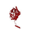
 Components Components | Tetrathionate hydrolase | ||||||
 Keywords Keywords |  HYDROLASE / Tetrathionate HYDROLASE / Tetrathionate | ||||||
| Function / homology |  Function and homology informationHydrolases; Acting on sulfur-sulfur bonds / hydrolase activity / plasma membrane Function and homology informationHydrolases; Acting on sulfur-sulfur bonds / hydrolase activity / plasma membraneSimilarity search - Function | ||||||
| Biological species |  Acidithiobacillus ferrooxidans (bacteria) Acidithiobacillus ferrooxidans (bacteria) | ||||||
| Method | X-RAY DIFFRACTION / SYNCHROTRON / SAD / Resolution: 1.95004814203 Å | ||||||
 Authors Authors | Tamada, T. / Hirano, Y. | ||||||
 Citation Citation | Journal: Protein Sci. / Year: 2020 Title: Reaction mechanism of tetrathionate hydrolysis based on the crystal structure of tetrathionate hydrolase from Acidithiobacillus ferrooxidans. Authors: Kanao, T. / Hase, N. / Nakayama, H. / Yoshida, K. / Nishiura, K. / Kosaka, M. / Kamimura, K. / Hirano, Y. / Tamada, T. | ||||||
| History |
|
- Structure visualization
| Structure viewer | Molecule: MolmilJmol/JSmol |
|---|
- Downloads & links
-Download
| PDBx/mmCIF format | 6l8a.cif.gz | 642.2 KB | Display | PDBx/mmCIF format |
|---|---|---|---|---|
| PDB format | pdb6l8a.ent.gz | 423.3 KB | Display | PDB format |
| PDBx/mmJSON format | 6l8a.json.gz | Tree view | PDBx/mmJSON format | |
| Others |  Other downloads Other downloads |
-Validation report
| Arichive directory | https://data.pdbj.org/pub/pdb/validation_reports/l8/6l8aftp://data.pdbj.org/pub/pdb/validation_reports/l8/6l8a | HTTPS FTP |
|---|
-Related structure data


-Links
 PDBj
PDBj

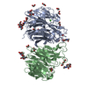
- Assembly
| Deposited unit | 
| ||||||||||
|---|---|---|---|---|---|---|---|---|---|---|---|
| 1 | 
| ||||||||||
| 2 | 
| ||||||||||
| 3 | 
| ||||||||||
| Unit cell |
|
-Components
| #1: Protein | Mass: 50047.230 Da / Num. of mol.: 6 Source method: isolated from a genetically manipulated source Source: (gene. exp.) Acidithiobacillus ferrooxidans (bacteria)Production host: Escherichia coli (E. coli)References: UniProt: B7J3C9, Hydrolases; Acting on sulfur-sulfur bonds#2: Chemical | ChemComp-SO4 / Sulfate  Mass: 96.063 Da / Num. of mol.: 30 / Source method: obtained synthetically / Formula: SO4 Mass: 96.063 Da / Num. of mol.: 30 / Source method: obtained synthetically / Formula: SO4#3: Chemical | Β-Alanine  Type: peptide-like / Mass: 89.093 Da / Num. of mol.: 3 / Source method: isolated from a natural source / Formula: C3H7NO2 Type: peptide-like / Mass: 89.093 Da / Num. of mol.: 3 / Source method: isolated from a natural source / Formula: C3H7NO2#4: Chemical | Glycine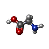  Type: peptide linking / Mass: 75.067 Da / Num. of mol.: 2 / Source method: obtained synthetically / Formula: C2H5NO2 Type: peptide linking / Mass: 75.067 Da / Num. of mol.: 2 / Source method: obtained synthetically / Formula: C2H5NO2#5: Water | ChemComp-HOH / | Water Mass: 18.015 Da / Num. of mol.: 697 / Source method: obtained synthetically / Formula: H2O Mass: 18.015 Da / Num. of mol.: 697 / Source method: obtained synthetically / Formula: H2OHas ligand of interest | N | |
|---|
-Experimental details
-Experiment
| Experiment | Method: X-RAY DIFFRACTION / Number of used crystals: 1 |
|---|
- Sample preparation
| Crystal | Density Matthews: 1.93 Å3/Da / Density % sol: 36.42 % |
|---|---|
| Crystal grow | Temperature: 291 K / Method: vapor diffusion, hanging drop / Details: sodium chloride, glycine, PEG 1000 |
-Data collection
| Diffraction | Mean temperature: 100 K / Serial crystal experiment: N |
|---|---|
| Diffraction source | Source: SYNCHROTRON / Site: SPring-8  / Beamline: BL41XU / Wavelength: 1 Å / Beamline: BL41XU / Wavelength: 1 Å |
| Detector | Type: MARMOSAIC 225 mm CCD / Detector: CCD / Date: Sep 27, 2012 |
| Radiation | Protocol: SINGLE WAVELENGTH / Monochromatic (M) / Laue (L): M / Scattering type: x-ray |
| Radiation wavelength | Wavelength: 1 Å / Relative weight: 1 |
| Reflection | Resolution: 1.95→42.4 Å / Num. obs: 156219 / % possible obs: 99.7 % / Redundancy: 3.8 % / Biso Wilson estimate: 25.7759592713 Å2 / CC1/2: 0.997 / Rrim(I) all: 0.109 / Net I/av σ(I): 10.3 / Net I/σ(I): 20.7 |
| Reflection shell | Resolution: 1.95→2.02 Å / Redundancy: 3.8 % / Mean I/σ(I) obs: 2.9 / Num. unique obs: 15660 / CC1/2: 0.875 / Rrim(I) all: 0.563 / % possible all: 99.9 |
- Processing
| Software |
| |||||||||||||||||||||||||||||||||||||||||||||||||||||||||||||||||||||||||||||||||||||||||||||||||||||||||||||||||||||||||||||||||||||||||||||||||||||||||||||||||||||||||||||||||||||||||||||||||||||||||||||||||||||||||
|---|---|---|---|---|---|---|---|---|---|---|---|---|---|---|---|---|---|---|---|---|---|---|---|---|---|---|---|---|---|---|---|---|---|---|---|---|---|---|---|---|---|---|---|---|---|---|---|---|---|---|---|---|---|---|---|---|---|---|---|---|---|---|---|---|---|---|---|---|---|---|---|---|---|---|---|---|---|---|---|---|---|---|---|---|---|---|---|---|---|---|---|---|---|---|---|---|---|---|---|---|---|---|---|---|---|---|---|---|---|---|---|---|---|---|---|---|---|---|---|---|---|---|---|---|---|---|---|---|---|---|---|---|---|---|---|---|---|---|---|---|---|---|---|---|---|---|---|---|---|---|---|---|---|---|---|---|---|---|---|---|---|---|---|---|---|---|---|---|---|---|---|---|---|---|---|---|---|---|---|---|---|---|---|---|---|---|---|---|---|---|---|---|---|---|---|---|---|---|---|---|---|---|---|---|---|---|---|---|---|---|---|---|---|---|---|---|---|---|
| Refinement | Method to determine structure: SAD / Resolution: 1.95004814203→42.3647721231 Å / SU ML: 0.227474103365 / Cross valid method: FREE R-VALUE / σ(F): 1.96795806727 / Phase error: 23.0146924341 Stereochemistry target values: GeoStd + Monomer Library + CDL v1.2
| |||||||||||||||||||||||||||||||||||||||||||||||||||||||||||||||||||||||||||||||||||||||||||||||||||||||||||||||||||||||||||||||||||||||||||||||||||||||||||||||||||||||||||||||||||||||||||||||||||||||||||||||||||||||||
| Solvent computation | Shrinkage radii: 0.9 Å / VDW probe radii: 1.11 Å / Solvent model: FLAT BULK SOLVENT MODEL | |||||||||||||||||||||||||||||||||||||||||||||||||||||||||||||||||||||||||||||||||||||||||||||||||||||||||||||||||||||||||||||||||||||||||||||||||||||||||||||||||||||||||||||||||||||||||||||||||||||||||||||||||||||||||
| Displacement parameters | Biso mean: 25.7975605035 Å2 | |||||||||||||||||||||||||||||||||||||||||||||||||||||||||||||||||||||||||||||||||||||||||||||||||||||||||||||||||||||||||||||||||||||||||||||||||||||||||||||||||||||||||||||||||||||||||||||||||||||||||||||||||||||||||
| Refinement step | Cycle: LAST / Resolution: 1.95004814203→42.3647721231 Å
| |||||||||||||||||||||||||||||||||||||||||||||||||||||||||||||||||||||||||||||||||||||||||||||||||||||||||||||||||||||||||||||||||||||||||||||||||||||||||||||||||||||||||||||||||||||||||||||||||||||||||||||||||||||||||
| Refine LS restraints |
| |||||||||||||||||||||||||||||||||||||||||||||||||||||||||||||||||||||||||||||||||||||||||||||||||||||||||||||||||||||||||||||||||||||||||||||||||||||||||||||||||||||||||||||||||||||||||||||||||||||||||||||||||||||||||
| LS refinement shell |
|
