 Movie
Movie Controller

[English] 日本語
 Yorodumi
- PDB-6kjo: The microtubule-binding domains of yeast cytoplasmic dynein in th... -
+ Open data

- Basic information
| Entry | Database: PDB / ID: 6kjo | |||||||||||||||
|---|---|---|---|---|---|---|---|---|---|---|---|---|---|---|---|---|
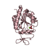
| Title | The microtubule-binding domains of yeast cytoplasmic dynein in the low affinity state | |||||||||||||||
 Components Components | Dynein heavy chain, cytoplasmic | |||||||||||||||
 Keywords Keywords |  MOTOR PROTEIN / Microtubule / Dynein / disulfide bond / low affinity MOTOR PROTEIN / Microtubule / Dynein / disulfide bond / low affinity | |||||||||||||||
| Function / homology |  Function and homology informationkaryogamy / establishment of mitotic spindle localization / astral microtubule / nuclear migration along microtubule / minus-end-directed microtubule motor activity / cytoplasmic dynein complex / dynein light intermediate chain binding / spindle pole body / nuclear migration / dynein intermediate chain binding ...karyogamy / establishment of mitotic spindle localization / astral microtubule / nuclear migration along microtubule / minus-end-directed microtubule motor activity / cytoplasmic dynein complex / dynein light intermediate chain binding / spindle pole body / nuclear migration / dynein intermediate chain binding / mitotic sister chromatid segregation / establishment of mitotic spindle orientation / cytoplasmic microtubule / cytoplasmic microtubule organization / Neutrophil degranulation / mitotic spindle organization / cell cortex / ATP hydrolysis activity / ATP binding / cytoplasm Function and homology informationkaryogamy / establishment of mitotic spindle localization / astral microtubule / nuclear migration along microtubule / minus-end-directed microtubule motor activity / cytoplasmic dynein complex / dynein light intermediate chain binding / spindle pole body / nuclear migration / dynein intermediate chain binding ...karyogamy / establishment of mitotic spindle localization / astral microtubule / nuclear migration along microtubule / minus-end-directed microtubule motor activity / cytoplasmic dynein complex / dynein light intermediate chain binding / spindle pole body / nuclear migration / dynein intermediate chain binding / mitotic sister chromatid segregation / establishment of mitotic spindle orientation / cytoplasmic microtubule / cytoplasmic microtubule organization / Neutrophil degranulation / mitotic spindle organization / cell cortex / ATP hydrolysis activity / ATP binding / cytoplasmSimilarity search - Function | |||||||||||||||
| Biological species |  Saccharomyces cerevisiae (brewer's yeast) Saccharomyces cerevisiae (brewer's yeast) | |||||||||||||||
| Method | SOLUTION NMR / simulated annealing | |||||||||||||||
 Authors Authors | Nishida, N. / Komori, Y. / Takarada, O. / Watanabe, A. / Tamura, S. / Kubo, S. / Shimada, I. / Kikkawa, M. | |||||||||||||||
| Funding support |  Japan, 4items Japan, 4items
| |||||||||||||||
 Citation Citation | Journal: Nat Commun / Year: 2020 Title: Structural basis for two-way communication between dynein and microtubules. Authors: Noritaka Nishida / Yuta Komori / Osamu Takarada / Atsushi Watanabe / Satoko Tamura / Satoshi Kubo / Ichio Shimada / Masahide Kikkawa / Abstract: The movements of cytoplasmic dynein on microtubule (MT) tracks is achieved by two-way communication between the microtubule-binding domain (MTBD) and the ATPase domain via a coiled-coil stalk, but ...The movements of cytoplasmic dynein on microtubule (MT) tracks is achieved by two-way communication between the microtubule-binding domain (MTBD) and the ATPase domain via a coiled-coil stalk, but the structural basis of this communication remains elusive. Here, we regulate MTBD either in high-affinity or low-affinity states by introducing a disulfide bond to the stalk and analyze the resulting structures by NMR and cryo-EM. In the MT-unbound state, the affinity changes of MTBD are achieved by sliding of the stalk α-helix by a half-turn, which suggests that structural changes propagate from the ATPase-domain to MTBD. In addition, MT binding induces further sliding of the stalk α-helix even without the disulfide bond, suggesting how the MT-induced conformational changes propagate toward the ATPase domain. Based on differences in the MT-binding surface between the high- and low-affinity states, we propose a potential mechanism for the directional bias of dynein movement on MT tracks. | |||||||||||||||
| History |
|
- Structure visualization
| Structure viewer | Molecule: MolmilJmol/JSmol |
|---|
- Downloads & links
-Download
| PDBx/mmCIF format | 6kjo.cif.gz | 454.9 KB | Display | PDBx/mmCIF format |
|---|---|---|---|---|
| PDB format | pdb6kjo.ent.gz | 382.3 KB | Display | PDB format |
| PDBx/mmJSON format | 6kjo.json.gz | Tree view | PDBx/mmJSON format | |
| Others |  Other downloads Other downloads |
-Validation report
| Arichive directory | https://data.pdbj.org/pub/pdb/validation_reports/kj/6kjoftp://data.pdbj.org/pub/pdb/validation_reports/kj/6kjo | HTTPS FTP |
|---|
-Related structure data
| Related structure data | 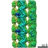 9996C 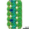 9997C  6kioC 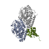 6kiqC 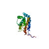 6kjnC C: citing same article ( |
|---|---|
| Similar structure data | |
| Other databases |


-Links
 PDBj
PDBj


- Assembly
| Deposited unit | 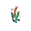
| |||||||||
|---|---|---|---|---|---|---|---|---|---|---|
| 1 |
| |||||||||
| NMR ensembles |
|
-Components
| #1: Protein | Mass: 16525.951 Da / Num. of mol.: 1 / Fragment: The microtubule-binding domain / Mutation: S3097C, V3222C Source method: isolated from a genetically manipulated source Source: (gene. exp.) Saccharomyces cerevisiae (strain ATCC 204508 / S288c) (yeast)Strain: ATCC 204508 / S288c / Gene: DYN1, DHC1, YKR054C / Plasmid: pET-15b / Cell (production host): BL21(DE3) / Production host:  Escherichia coli (E. coli) / References: UniProt: P36022 Escherichia coli (E. coli) / References: UniProt: P36022 |
|---|
-Experimental details
-Experiment
| Experiment | Method: SOLUTION NMR | ||||||||||||||||||||||||||||||||||||||||||||||||||||||||||||||||||||||||
|---|---|---|---|---|---|---|---|---|---|---|---|---|---|---|---|---|---|---|---|---|---|---|---|---|---|---|---|---|---|---|---|---|---|---|---|---|---|---|---|---|---|---|---|---|---|---|---|---|---|---|---|---|---|---|---|---|---|---|---|---|---|---|---|---|---|---|---|---|---|---|---|---|---|
| NMR experiment |
|
- Sample preparation
| Details |
| |||||||||||||||
|---|---|---|---|---|---|---|---|---|---|---|---|---|---|---|---|---|
| Sample |
| |||||||||||||||
| Sample conditions | Ionic strength: 200 mM / Label: conditions_1 / pH: 7.0 / Pressure: 1 atm / Temperature: 298 K |
-NMR measurement
| NMR spectrometer | Type: Bruker AVANCE III / Manufacturer: Bruker / Model: AVANCE III / Field strength: 800 MHz |
|---|
- Processing
| NMR software |
| ||||||||||||
|---|---|---|---|---|---|---|---|---|---|---|---|---|---|
| Refinement | Method: simulated annealing / Software ordinal: 2 | ||||||||||||
| NMR representative | Selection criteria: lowest energy | ||||||||||||
| NMR ensemble | Conformer selection criteria: structures with the lowest energy Conformers calculated total number: 200 / Conformers submitted total number: 10 |
