 Movie
Movie Controller

[English] 日本語
 Yorodumi
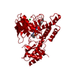
- PDB-6kg1: NifS from Helicobacter pylori, soaked with L-cysteine for 180 sec -
+ Open data

- Basic information
| Entry | Database: PDB / ID: 6kg1 | ||||||
|---|---|---|---|---|---|---|---|
| Title | NifS from Helicobacter pylori, soaked with L-cysteine for 180 sec | ||||||
 Components Components | Cysteine desulfurase IscS | ||||||
 Keywords Keywords |  BIOSYNTHETIC PROTEIN / iron / sulfur / cysteine desulfurase / reaction intermediate BIOSYNTHETIC PROTEIN / iron / sulfur / cysteine desulfurase / reaction intermediate | ||||||
| Function / homology |  Function and homology informationcysteine desulfurase / cysteine desulfurase activity / cysteine metabolic process / [2Fe-2S] cluster assembly / 2 iron, 2 sulfur cluster binding / pyridoxal phosphate binding / metal ion binding / cytoplasm Function and homology informationcysteine desulfurase / cysteine desulfurase activity / cysteine metabolic process / [2Fe-2S] cluster assembly / 2 iron, 2 sulfur cluster binding / pyridoxal phosphate binding / metal ion binding / cytoplasmSimilarity search - Function | ||||||
| Biological species |   Helicobacter pylori (bacteria) Helicobacter pylori (bacteria) | ||||||
| Method | X-RAY DIFFRACTION / SYNCHROTRON / MOLECULAR REPLACEMENT / Resolution: 2.7 Å | ||||||
 Authors Authors | Nakamura, R. / Takahashi, Y. / Fujishiro, T. | ||||||
| Funding support |  Japan, 1items Japan, 1items
| ||||||
 Citation Citation | Journal: Febs J. / Year: 2020 Title: Snapshots of PLP-substrate and PLP-product external aldimines as intermediates in two types of cysteine desulfurase enzymes. Authors: Nakamura, R. / Hikita, M. / Ogawa, S. / Takahashi, Y. / Fujishiro, T. | ||||||
| History |
|
- Structure visualization
| Structure viewer | Molecule: MolmilJmol/JSmol |
|---|
- Downloads & links
-Download
| PDBx/mmCIF format | 6kg1.cif.gz | 161 KB | Display | PDBx/mmCIF format |
|---|---|---|---|---|
| PDB format | pdb6kg1.ent.gz | 125.5 KB | Display | PDB format |
| PDBx/mmJSON format | 6kg1.json.gz | Tree view | PDBx/mmJSON format | |
| Others |  Other downloads Other downloads |
-Validation report
| Arichive directory | https://data.pdbj.org/pub/pdb/validation_reports/kg/6kg1ftp://data.pdbj.org/pub/pdb/validation_reports/kg/6kg1 | HTTPS FTP |
|---|
-Related structure data
| Related structure data |  5wt2SC  5wt4C  5zs9C  5zskC  5zsoC  5zspC  5zstC  6kfzC  6kg0C S: Starting model for refinement C: citing same article ( |
|---|---|
| Similar structure data |


-Links
 PDBj
PDBj
- Assembly
| Deposited unit | 
| ||||||||
|---|---|---|---|---|---|---|---|---|---|
| 1 | 
| ||||||||
| Unit cell |
|
-Components
| #1: Protein | Mass: 44125.230 Da / Num. of mol.: 1 / Mutation: L2V, K138R Source method: isolated from a genetically manipulated source Source: (gene. exp.) Helicobacter pylori (strain ATCC 700392 / 26695) (bacteria)Strain: ATCC 700392 / 26695 / Gene: iscS, HP_0220 / Production host: Escherichia coli BL21(DE3) (bacteria) / Variant (production host): C41 / References: UniProt: O25008, cysteine desulfurase |
|---|---|
| #2: Chemical | ChemComp-IPA / Isopropyl alcohol  Mass: 60.095 Da / Num. of mol.: 1 / Source method: obtained synthetically / Formula: C3H8O / Comment: alkaloid*YM Mass: 60.095 Da / Num. of mol.: 1 / Source method: obtained synthetically / Formula: C3H8O / Comment: alkaloid*YM |
| #3: Chemical | ChemComp-CL / Chloride  Mass: 35.453 Da / Num. of mol.: 1 / Source method: obtained synthetically / Formula: Cl Mass: 35.453 Da / Num. of mol.: 1 / Source method: obtained synthetically / Formula: Cl |
| #4: Chemical | ChemComp-PDA /   Mass: 320.236 Da / Num. of mol.: 1 / Source method: obtained synthetically / Formula: C11H17N2O7P Mass: 320.236 Da / Num. of mol.: 1 / Source method: obtained synthetically / Formula: C11H17N2O7P |
| #5: Water | ChemComp-HOH / Water Mass: 18.015 Da / Num. of mol.: 8 / Source method: isolated from a natural source / Formula: H2O Mass: 18.015 Da / Num. of mol.: 8 / Source method: isolated from a natural source / Formula: H2O |
| Has ligand of interest | N |
-Experimental details
-Experiment
| Experiment | Method: X-RAY DIFFRACTION / Number of used crystals: 1 |
|---|
- Sample preparation
| Crystal | Density Matthews: 4 Å3/Da / Density % sol: 69.22 % |
|---|---|
| Crystal grow | Temperature: 293 K / Method: vapor diffusion, sitting drop / pH: 7.5 Details: 17% (w/v) PEG 4000, 8.5% (v/v) 2-propanol, 15% (v/v) glycerol, 85mM HEPES-NaOH |
-Data collection
| Diffraction | Mean temperature: 100 K / Serial crystal experiment: N | ||||||||||||||||||||||||||||||||||||||||||||||||||||||||||||||||||||||||||||||||
|---|---|---|---|---|---|---|---|---|---|---|---|---|---|---|---|---|---|---|---|---|---|---|---|---|---|---|---|---|---|---|---|---|---|---|---|---|---|---|---|---|---|---|---|---|---|---|---|---|---|---|---|---|---|---|---|---|---|---|---|---|---|---|---|---|---|---|---|---|---|---|---|---|---|---|---|---|---|---|---|---|---|
| Diffraction source | Source: SYNCHROTRON / Site: Photon Factory / Beamline: BL-17A / Wavelength: 0.98 Å | ||||||||||||||||||||||||||||||||||||||||||||||||||||||||||||||||||||||||||||||||
| Detector | Type: DECTRIS EIGER X 16M / Detector: PIXEL / Date: Jun 29, 2019 / Details: Si(111) double crystal monochromator | ||||||||||||||||||||||||||||||||||||||||||||||||||||||||||||||||||||||||||||||||
| Radiation | Monochromator: Si(111) double crystal monochromator / Protocol: SINGLE WAVELENGTH / Monochromatic (M) / Laue (L): M / Scattering type: x-ray | ||||||||||||||||||||||||||||||||||||||||||||||||||||||||||||||||||||||||||||||||
| Radiation wavelength | Wavelength: 0.98 Å / Relative weight: 1 | ||||||||||||||||||||||||||||||||||||||||||||||||||||||||||||||||||||||||||||||||
| Reflection | Resolution: 2.7→49.18 Å / Num. obs: 20267 / % possible obs: 100 % / Redundancy: 7.086 % / Biso Wilson estimate: 73.219 Å2 / CC1/2: 0.998 / Rmerge(I) obs: 0.083 / Rrim(I) all: 0.09 / Χ2: 1.019 / Net I/σ(I): 14.18 / Num. measured all: 265780 / Scaling rejects: 7 | ||||||||||||||||||||||||||||||||||||||||||||||||||||||||||||||||||||||||||||||||
| Reflection shell | Diffraction-ID: 1
|
- Processing
| Software |
| ||||||||||||||||||||||||||||||||||||||||||||||||||||||||||||||||||||||||||||||||||||||||||||||||||||
|---|---|---|---|---|---|---|---|---|---|---|---|---|---|---|---|---|---|---|---|---|---|---|---|---|---|---|---|---|---|---|---|---|---|---|---|---|---|---|---|---|---|---|---|---|---|---|---|---|---|---|---|---|---|---|---|---|---|---|---|---|---|---|---|---|---|---|---|---|---|---|---|---|---|---|---|---|---|---|---|---|---|---|---|---|---|---|---|---|---|---|---|---|---|---|---|---|---|---|---|---|---|
| Refinement | Method to determine structure: MOLECULAR REPLACEMENT Starting model: 5WT2 Resolution: 2.7→49.18 Å / Cor.coef. Fo:Fc: 0.961 / Cor.coef. Fo:Fc free: 0.934 / SU B: 22.207 / SU ML: 0.205 / Cross valid method: THROUGHOUT / σ(F): 0 / ESU R: 0.294 / ESU R Free: 0.243 / Details: U VALUES : WITH TLS ADDED
| ||||||||||||||||||||||||||||||||||||||||||||||||||||||||||||||||||||||||||||||||||||||||||||||||||||
| Solvent computation | Ion probe radii: 0.8 Å / Shrinkage radii: 0.8 Å / VDW probe radii: 1.2 Å | ||||||||||||||||||||||||||||||||||||||||||||||||||||||||||||||||||||||||||||||||||||||||||||||||||||
| Displacement parameters | Biso max: 204.99 Å2 / Biso mean: 92.068 Å2 / Biso min: 38.4 Å2
| ||||||||||||||||||||||||||||||||||||||||||||||||||||||||||||||||||||||||||||||||||||||||||||||||||||
| Refinement step | Cycle: final / Resolution: 2.7→49.18 Å
| ||||||||||||||||||||||||||||||||||||||||||||||||||||||||||||||||||||||||||||||||||||||||||||||||||||
| Refine LS restraints |
| ||||||||||||||||||||||||||||||||||||||||||||||||||||||||||||||||||||||||||||||||||||||||||||||||||||
| LS refinement shell | Resolution: 2.7→2.77 Å / Rfactor Rfree error: 0 / Total num. of bins used: 20
| ||||||||||||||||||||||||||||||||||||||||||||||||||||||||||||||||||||||||||||||||||||||||||||||||||||
| Refinement TLS params. | Method: refined / Refine-ID: X-RAY DIFFRACTION
| ||||||||||||||||||||||||||||||||||||||||||||||||||||||||||||||||||||||||||||||||||||||||||||||||||||
| Refinement TLS group |
|
