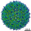
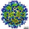
Journal: Cell Rep / Year: 2019 Title: Structural Basis for Neutralization and Protection by a Zika Virus-Specific Human Antibody. Authors: Lin Wang / Ruoke Wang / Lei Wang / Haijing Ben / Lei Yu / Fei Gao / Xuanling Shi / Chibiao Yin / Fuchun Zhang / Ye Xiang / Linqi Zhang / Abstract: We previously reported a human monoclonal antibody, ZK2B10, capable of protection against Zika virus (ZIKV) infection and microcephaly in developing mouse embryos. Here, we report the structural ...We previously reported a human monoclonal antibody, ZK2B10, capable of protection against Zika virus (ZIKV) infection and microcephaly in developing mouse embryos. Here, we report the structural features and mechanism of action of ZK2B10. The crystal structure at a resolution of 2.32 Å revealed that the epitope is located on the lateral ridge of DIII of the envelope glycoprotein. Cryo-EM structure with mature ZIKV showed that the antibody binds to DIIIs around the icosahedral 2-fold, 3-fold, and 5-fold axes, a distinct feature compared to those reported for DIII-specific antibodies. The binding of ZK2B10 to ZIKV has no detectable effect on viral attachment to target cells or on conformational changes of the E glycoprotein in the acidic environment, suggesting that ZK2B10 functions at steps between the formation of the fusion intermediate and membrane fusion. These results provide structural and mechanistic insights into how ZK2B10 mediates protection against ZIKV infection.
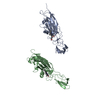
H: heavy chain of Fab ZK2B10 L: light chain of Fab ZK2B10 E: Genome polyprotein K: heavy chain of Fab ZK2B10 I: light chain of Fab ZK2B10 F: Genome polyprotein
Mass: 25383.305 Da / Num. of mol.: 2 Source method: isolated from a genetically manipulated source Source: (gene. exp.) Homo sapiens (human) / Production host: Homo sapiens (human)
#2: Antibody
lightchainofFabZK2B10
Mass: 22782.035 Da / Num. of mol.: 2 Source method: isolated from a genetically manipulated source Source: (gene. exp.) Homo sapiens (human) / Production host: Homo sapiens (human)
#3: Protein
Genomepolyprotein
Mass: 10989.564 Da / Num. of mol.: 2 Source method: isolated from a genetically manipulated source Source: (gene. exp.) Zika virus / Production host: Escherichia coli K-12 (bacteria) / Strain (production host): K-12 / References: UniProt: A0A2Z2ESG6
In the structure databanks used in Yorodumi, some data are registered as the other names, "COVID-19 virus" and "2019-nCoV". Here are the details of the virus and the list of structure data.
Jan 31, 2019. EMDB accession codes are about to change! (news from PDBe EMDB page)
EMDB accession codes are about to change! (news from PDBe EMDB page)
The allocation of 4 digits for EMDB accession codes will soon come to an end. Whilst these codes will remain in use, new EMDB accession codes will include an additional digit and will expand incrementally as the available range of codes is exhausted. The current 4-digit format prefixed with “EMD-” (i.e. EMD-XXXX) will advance to a 5-digit format (i.e. EMD-XXXXX), and so on. It is currently estimated that the 4-digit codes will be depleted around Spring 2019, at which point the 5-digit format will come into force.
The EM Navigator/Yorodumi systems omit the EMD- prefix.
Related info.:Q: What is EMD? / ID/Accession-code notation in Yorodumi/EM Navigator
Yorodumi is a browser for structure data from EMDB, PDB, SASBDB, etc.
This page is also the successor to EM Navigator detail page, and also detail information page/front-end page for Omokage search.
The word "yorodu" (or yorozu) is an old Japanese word meaning "ten thousand". "mi" (miru) is to see.
Related info.:EMDB / PDB / SASBDB / Comparison of 3 databanks / Yorodumi Search / Aug 31, 2016. New EM Navigator & Yorodumi / Yorodumi Papers / Jmol/JSmol / Function and homology information / Changes in new EM Navigator and Yorodumi
 Movie
Movie Controller

 Yorodumi
 Open data
 Basic information
 Components
 Keywords
 ANTIVIRAL PROTEIN /
  Function and homology information

 Authors
 Citation
 Structure visualization
 Downloads & links
 Other downloads
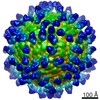


 PDBj
PDBj


 Assembly


 Mass: 18.015 Da / Num. of mol.: 580 / Source method: isolated from a natural source / Formula: H2O
 Sample preparation
 Processing