 Movie
Movie Controller

[English] 日本語
 Yorodumi
- PDB-6j1b: Photoswitchable fluorescent protein Gamillus, N150C/T204V double ... -
+ Open data

- Basic information
| Entry | Database: PDB / ID: 6j1b | |||||||||
|---|---|---|---|---|---|---|---|---|---|---|
| Title | Photoswitchable fluorescent protein Gamillus, N150C/T204V double mutant, on-state | |||||||||
 Components Components | Green fluorescent protein | |||||||||
 Keywords Keywords | FLUORESCENT PROTEIN / acid-tolerant / photoswitchable / N150C/T204V double mutant / trans-chromophore | |||||||||
| Function / homology |  Function and homology information Function and homology information | |||||||||
| Biological species |  Olindias (invertebrata) Olindias (invertebrata) | |||||||||
| Method | X-RAY DIFFRACTION / MOLECULAR REPLACEMENT / Resolution: 1.99 Å | |||||||||
 Authors Authors | Nakashima, R. / Shinoda, H. / Matsuda, T. / Nagai, T. | |||||||||
 Citation Citation | Journal: Cell Chem Biol / Year: 2019 Title: Acid-Tolerant Reversibly Switchable Green Fluorescent Protein for Super-resolution Imaging under Acidic Conditions. Authors: Shinoda, H. / Lu, K. / Nakashima, R. / Wazawa, T. / Noguchi, K. / Matsuda, T. / Nagai, T. | |||||||||
| History |
|
- Structure visualization
| Structure viewer | Molecule: MolmilJmol/JSmol |
|---|
- Downloads & links
-Download
| PDBx/mmCIF format | 6j1b.cif.gz | 68.3 KB | Display | PDBx/mmCIF format |
|---|---|---|---|---|
| PDB format | pdb6j1b.ent.gz | 47.4 KB | Display | PDB format |
| PDBx/mmJSON format | 6j1b.json.gz | Tree view | PDBx/mmJSON format | |
| Others |  Other downloads Other downloads |
-Validation report
| Arichive directory | https://data.pdbj.org/pub/pdb/validation_reports/j1/6j1bftp://data.pdbj.org/pub/pdb/validation_reports/j1/6j1b | HTTPS FTP |
|---|
-Related structure data
| Related structure data | 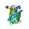 6j1aC  6j1cC  6jxfC  5y00S S: Starting model for refinement C: citing same article ( |
|---|---|
| Similar structure data |


-Links
 PDBj
PDBj

- Assembly
| Deposited unit | 
| ||||||||
|---|---|---|---|---|---|---|---|---|---|
| 1 |
| ||||||||
| Unit cell |
|
-Components
| #1: Protein | Mass: 30397.307 Da / Num. of mol.: 1 / Mutation: N150C, T204V Source method: isolated from a genetically manipulated source Source: (gene. exp.) Olindias (invertebrata) / Production host:  Escherichia coli (E. coli) / References: UniProt: A0A2Z5X7U3*PLUS Escherichia coli (E. coli) / References: UniProt: A0A2Z5X7U3*PLUS | ||||
|---|---|---|---|---|---|
| #2: Chemical | ChemComp-PO4 / Phosphate  Mass: 94.971 Da / Num. of mol.: 1 / Source method: obtained synthetically / Formula: PO4 Mass: 94.971 Da / Num. of mol.: 1 / Source method: obtained synthetically / Formula: PO4 | ||||
| #3: Chemical | ChemComp-GOL / Glycerol  Mass: 92.094 Da / Num. of mol.: 1 / Source method: obtained synthetically / Formula: C3H8O3 Mass: 92.094 Da / Num. of mol.: 1 / Source method: obtained synthetically / Formula: C3H8O3 | ||||
| #4: Chemical | Chloride  Mass: 35.453 Da / Num. of mol.: 2 / Source method: obtained synthetically / Formula: Cl Mass: 35.453 Da / Num. of mol.: 2 / Source method: obtained synthetically / Formula: Cl#5: Water | ChemComp-HOH / | Water Mass: 18.015 Da / Num. of mol.: 218 / Source method: isolated from a natural source / Formula: H2O Mass: 18.015 Da / Num. of mol.: 218 / Source method: isolated from a natural source / Formula: H2OSequence details | Residues 150CYS and 204VAL represent mutations (N150C/T204V). | |
-Experimental details
-Experiment
| Experiment | Method: X-RAY DIFFRACTION / Number of used crystals: 1 |
|---|
- Sample preparation
| Crystal | Density Matthews: 5.77 Å3/Da / Density % sol: 78.69 % |
|---|---|
| Crystal grow | Temperature: 298 K / Method: vapor diffusion, hanging drop / pH: 5 Details: mmonium phosphate, sodium citrate, sodium chloride, HEPES, pH5.0 |
-Data collection
| Diffraction | Mean temperature: 100 K / Serial crystal experiment: N | ||||||||||||||||||||||||||||||||||||||||||||||||||||||||||||||||||||||||||||||||||||||||||||||||||||
|---|---|---|---|---|---|---|---|---|---|---|---|---|---|---|---|---|---|---|---|---|---|---|---|---|---|---|---|---|---|---|---|---|---|---|---|---|---|---|---|---|---|---|---|---|---|---|---|---|---|---|---|---|---|---|---|---|---|---|---|---|---|---|---|---|---|---|---|---|---|---|---|---|---|---|---|---|---|---|---|---|---|---|---|---|---|---|---|---|---|---|---|---|---|---|---|---|---|---|---|---|---|
| Diffraction source | Source: ROTATING ANODE / Type: RIGAKU FR-E+ SUPERBRIGHT / Wavelength: 1.5418 Å | ||||||||||||||||||||||||||||||||||||||||||||||||||||||||||||||||||||||||||||||||||||||||||||||||||||
| Detector | Type: RIGAKU RAXIS IV++ / Detector: IMAGE PLATE / Date: Dec 11, 2018 | ||||||||||||||||||||||||||||||||||||||||||||||||||||||||||||||||||||||||||||||||||||||||||||||||||||
| Radiation | Protocol: SINGLE WAVELENGTH / Monochromatic (M) / Laue (L): M / Scattering type: x-ray | ||||||||||||||||||||||||||||||||||||||||||||||||||||||||||||||||||||||||||||||||||||||||||||||||||||
| Radiation wavelength | Wavelength: 1.5418 Å / Relative weight: 1 | ||||||||||||||||||||||||||||||||||||||||||||||||||||||||||||||||||||||||||||||||||||||||||||||||||||
| Reflection | Resolution: 1.99→46.619 Å / Num. obs: 47626 / % possible obs: 100 % / Redundancy: 14.402 % / Biso Wilson estimate: 26.014 Å2 / CC1/2: 0.999 / Rmerge(I) obs: 0.071 / Rrim(I) all: 0.073 / Χ2: 1.026 / Net I/σ(I): 27.64 / Num. measured all: 685896 | ||||||||||||||||||||||||||||||||||||||||||||||||||||||||||||||||||||||||||||||||||||||||||||||||||||
| Reflection shell | Diffraction-ID: 1
|
- Processing
| Software |
| ||||||||||||||||||||||||||||||||||||||||||||||||||||||||||||
|---|---|---|---|---|---|---|---|---|---|---|---|---|---|---|---|---|---|---|---|---|---|---|---|---|---|---|---|---|---|---|---|---|---|---|---|---|---|---|---|---|---|---|---|---|---|---|---|---|---|---|---|---|---|---|---|---|---|---|---|---|---|
| Refinement | Method to determine structure: MOLECULAR REPLACEMENT Starting model: 5Y00 Resolution: 1.99→46.62 Å / Cor.coef. Fo:Fc: 0.967 / Cor.coef. Fo:Fc free: 0.959 / SU B: 1.39 / SU ML: 0.039 / Cross valid method: THROUGHOUT / σ(F): 0 / ESU R: 0.068 / ESU R Free: 0.069 / Stereochemistry target values: MAXIMUM LIKELIHOOD Details: HYDROGENS HAVE BEEN ADDED IN THE RIDING POSITIONS U VALUES : REFINED INDIVIDUALLY
| ||||||||||||||||||||||||||||||||||||||||||||||||||||||||||||
| Solvent computation | Ion probe radii: 0.8 Å / Shrinkage radii: 0.8 Å / VDW probe radii: 1.2 Å / Solvent model: MASK | ||||||||||||||||||||||||||||||||||||||||||||||||||||||||||||
| Displacement parameters | Biso max: 98.64 Å2 / Biso mean: 22.005 Å2 / Biso min: 10.9 Å2
| ||||||||||||||||||||||||||||||||||||||||||||||||||||||||||||
| Refinement step | Cycle: final / Resolution: 1.99→46.62 Å
| ||||||||||||||||||||||||||||||||||||||||||||||||||||||||||||
| Refine LS restraints |
| ||||||||||||||||||||||||||||||||||||||||||||||||||||||||||||
| LS refinement shell | Resolution: 1.995→2.047 Å / Rfactor Rfree error: 0 / Total num. of bins used: 20
|
