 Movie
Movie Controller


+ Open data

- Basic information
| Entry | Database: PDB / ID: 6fpt | ||||||
|---|---|---|---|---|---|---|---|
| Title | Crystal structure of Danio rerio Lin41 filamin-NHL domains | ||||||
 Components Components | E3 ubiquitin-protein ligase TRIM71 | ||||||
 Keywords Keywords |  RNA BINDING PROTEIN / post-transcriptional regulation RNA BINDING PROTEIN / post-transcriptional regulation | ||||||
| Function / homology |  Function and homology information Function and homology informationAntigen processing: Ubiquitination & Proteasome degradation / regulation of neural precursor cell proliferation / miRNA-mediated gene silencing by inhibition of translation / neural tube development / miRNA binding / fibroblast growth factor receptor signaling pathway / protein autoubiquitination / stem cell proliferation / P-body / RING-type E3 ubiquitin transferase ...Antigen processing: Ubiquitination & Proteasome degradation / regulation of neural precursor cell proliferation / miRNA-mediated gene silencing by inhibition of translation / neural tube development / miRNA binding / fibroblast growth factor receptor signaling pathway / protein autoubiquitination / stem cell proliferation / P-body / RING-type E3 ubiquitin transferase / G1/S transition of mitotic cell cycle / protein polyubiquitination / ubiquitin-protein transferase activity / ubiquitin protein ligase activity / proteasome-mediated ubiquitin-dependent protein catabolic process / zinc ion bindingSimilarity search - Function | ||||||
| Biological species |  Danio rerio (zebrafish) Danio rerio (zebrafish) | ||||||
| Method | X-RAY DIFFRACTION / SYNCHROTRON / MOLECULAR REPLACEMENT / Resolution: 2.6 Å | ||||||
 Authors Authors | Kumari, P. / Aeschimann, F. / Gaidatzis, D. / Keusch, J.J. / Ghosh, P. / Neagu, A. / Pachulska-Wieczorek, K. / Bujnicki, J.M. / Gut, H. / Grosshans, H. / Ciosk, R. | ||||||
 Citation Citation | Journal: Nat Commun / Year: 2018 Title: Evolutionary plasticity of the NHL domain underlies distinct solutions to RNA recognition. Authors: Kumari, P. / Aeschimann, F. / Gaidatzis, D. / Keusch, J.J. / Ghosh, P. / Neagu, A. / Pachulska-Wieczorek, K. / Bujnicki, J.M. / Gut, H. / Grosshans, H. / Ciosk, R. | ||||||
| History |
|
- Structure visualization
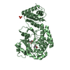
| Structure viewer | Molecule: MolmilJmol/JSmol |
|---|
- Downloads & links
-Download
| PDBx/mmCIF format | 6fpt.cif.gz | 307 KB | Display | PDBx/mmCIF format |
|---|---|---|---|---|
| PDB format | pdb6fpt.ent.gz | 250 KB | Display | PDB format |
| PDBx/mmJSON format | 6fpt.json.gz | Tree view | PDBx/mmJSON format | |
| Others |  Other downloads Other downloads |
-Validation report
| Arichive directory | https://data.pdbj.org/pub/pdb/validation_reports/fp/6fptftp://data.pdbj.org/pub/pdb/validation_reports/fp/6fpt | HTTPS FTP |
|---|
-Related structure data
| Related structure data |  6fq3C  6fqlC  1q7fS  4umgS S: Starting model for refinement C: citing same article ( |
|---|---|
| Similar structure data |


-Links
 PDBj
PDBj


- Assembly
| Deposited unit | 
| ||||||||
|---|---|---|---|---|---|---|---|---|---|
| 1 | 
| ||||||||
| 2 | 
| ||||||||
| Unit cell |
|
-Components
| #1: Protein | Mass: 44624.820 Da / Num. of mol.: 2 Source method: isolated from a genetically manipulated source Details: Fragment encompassing the filamin-NHL domain (residues 435-824) Source: (gene. exp.) Danio rerio (zebrafish) / Gene: trim71, lin41 / Cell line (production host): Sf9 / Production host:   Spodoptera frugiperda (fall armyworm) Spodoptera frugiperda (fall armyworm)References: UniProt: E7FAM5, RING-type E3 ubiquitin transferase #2: Water | ChemComp-HOH / | Water Mass: 18.015 Da / Num. of mol.: 215 / Source method: isolated from a natural source / Formula: H2O Mass: 18.015 Da / Num. of mol.: 215 / Source method: isolated from a natural source / Formula: H2O |
|---|
-Experimental details
-Experiment
| Experiment | Method: X-RAY DIFFRACTION / Number of used crystals: 1 |
|---|
- Sample preparation
| Crystal | Density Matthews: 2.19 Å3/Da / Density % sol: 43.79 % |
|---|---|
| Crystal grow | Temperature: 293 K / Method: vapor diffusion, sitting drop / pH: 7 Details: 10% PEG 8000 0.2 M magnesium chloride 0.1 M Tris pH 7.0 0.1 M trisodium citrate |
-Data collection
| Diffraction | Mean temperature: 100 K |
|---|---|
| Diffraction source | Source: SYNCHROTRON / Site: SLS  / Beamline: X06DA / Wavelength: 1.26 Å / Beamline: X06DA / Wavelength: 1.26 Å |
| Detector | Type: DECTRIS PILATUS 2M / Detector: PIXEL / Date: Jul 15, 2015 |
| Radiation | Protocol: SINGLE WAVELENGTH / Monochromatic (M) / Laue (L): M / Scattering type: x-ray |
| Radiation wavelength | Wavelength: 1.26 Å / Relative weight: 1 |
| Reflection | Resolution: 2.6→50 Å / Num. obs: 42647 / % possible obs: 91.6 % / Redundancy: 1.9 % / Biso Wilson estimate: 45.33 Å2 / CC1/2: 0.987 / Rsym value: 0.124 / Net I/σ(I): 5.28 |
| Reflection shell | Resolution: 2.6→2.67 Å / Redundancy: 1.8 % / Num. unique obs: 3006 / CC1/2: 0.779 / Rsym value: 0.491 / % possible all: 86.8 |
- Processing
| Software |
| ||||||||||||||||||||||||||||||||||||||||||||||||||||||||||||||||||||||||||||||||||||||||||||||||||||||||||||||||||
|---|---|---|---|---|---|---|---|---|---|---|---|---|---|---|---|---|---|---|---|---|---|---|---|---|---|---|---|---|---|---|---|---|---|---|---|---|---|---|---|---|---|---|---|---|---|---|---|---|---|---|---|---|---|---|---|---|---|---|---|---|---|---|---|---|---|---|---|---|---|---|---|---|---|---|---|---|---|---|---|---|---|---|---|---|---|---|---|---|---|---|---|---|---|---|---|---|---|---|---|---|---|---|---|---|---|---|---|---|---|---|---|---|---|---|---|
| Refinement | Method to determine structure: MOLECULAR REPLACEMENT Starting model: Homology models based on PDB IDs 4UMG and 1Q7F Resolution: 2.6→46.43 Å / Cor.coef. Fo:Fc: 0.9394 / Cor.coef. Fo:Fc free: 0.8825 / Cross valid method: THROUGHOUT / σ(F): 0 / SU Rfree Blow DPI: 0.329
| ||||||||||||||||||||||||||||||||||||||||||||||||||||||||||||||||||||||||||||||||||||||||||||||||||||||||||||||||||
| Displacement parameters | Biso mean: 30.59 Å2
| ||||||||||||||||||||||||||||||||||||||||||||||||||||||||||||||||||||||||||||||||||||||||||||||||||||||||||||||||||
| Refine analyze | Luzzati coordinate error obs: 0.309 Å | ||||||||||||||||||||||||||||||||||||||||||||||||||||||||||||||||||||||||||||||||||||||||||||||||||||||||||||||||||
| Refinement step | Cycle: 1 / Resolution: 2.6→46.43 Å
| ||||||||||||||||||||||||||||||||||||||||||||||||||||||||||||||||||||||||||||||||||||||||||||||||||||||||||||||||||
| Refine LS restraints |
| ||||||||||||||||||||||||||||||||||||||||||||||||||||||||||||||||||||||||||||||||||||||||||||||||||||||||||||||||||
| LS refinement shell | Resolution: 2.6→2.71 Å / Total num. of bins used: 12
| ||||||||||||||||||||||||||||||||||||||||||||||||||||||||||||||||||||||||||||||||||||||||||||||||||||||||||||||||||
| Refinement TLS params. | L22: 0 °2 / Method: refined / Refine-ID: X-RAY DIFFRACTION
| ||||||||||||||||||||||||||||||||||||||||||||||||||||||||||||||||||||||||||||||||||||||||||||||||||||||||||||||||||
| Refinement TLS group |
|
