 Movie
Movie Controller

[English] 日本語
 Yorodumi
- PDB-6fd9: Catalytic subunit HisG from Psychrobacter arcticus ATP phosphorib... -
+ Open data

- Basic information
| Entry | Database: PDB / ID: 6fd9 | ||||||
|---|---|---|---|---|---|---|---|
| Title | Catalytic subunit HisG from Psychrobacter arcticus ATP phosphoribosyltransferase (HisZG ATPPRT) in complex with AMP | ||||||
 Components Components | ATP phosphoribosyltransferase | ||||||
 Keywords Keywords | TRANSFERASE / phosphoribosyltransferase / HisG / catalytic / cold-adapted | ||||||
| Function / homology |  Function and homology informationATP phosphoribosyltransferase / ATP phosphoribosyltransferase activity / L-histidine biosynthetic process / ATP binding / cytoplasm Function and homology informationATP phosphoribosyltransferase / ATP phosphoribosyltransferase activity / L-histidine biosynthetic process / ATP binding / cytoplasmSimilarity search - Function | ||||||
| Biological species |  Psychrobacter arcticus (bacteria) Psychrobacter arcticus (bacteria) | ||||||
| Method | X-RAY DIFFRACTION / MOLECULAR REPLACEMENT / molecular replacement / Resolution: 2.2 Å | ||||||
 Authors Authors | Alphey, M.S. / Ge, Y. / Fisher, G. / Czekster, C.M. / Naismith, J.H. / da Silva, R.G. | ||||||
| Funding support |  United Kingdom, 1items United Kingdom, 1items
| ||||||
 Citation Citation | Journal: Acs Catalysis / Year: 2018 Title: Catalytic and Anticatalytic Snapshots of a Short-Form ATP Phosphoribosyltransferase Authors: Alphey, M.S. / Fisher, G. / Hirschi, J.S. / Stroek, R. / Ge, Y. / Gould, E.R. / Czekster, C.M. / Liu, H. / Florence, G.J. / Vetticatt, M.J. / Naismith, J.H. / da Silva, R.G. | ||||||
| History |
|
- Structure visualization
| Structure viewer | Molecule: MolmilJmol/JSmol |
|---|
- Downloads & links
-Download
| PDBx/mmCIF format | 6fd9.cif.gz | 57.5 KB | Display | PDBx/mmCIF format |
|---|---|---|---|---|
| PDB format | pdb6fd9.ent.gz | 39.6 KB | Display | PDB format |
| PDBx/mmJSON format | 6fd9.json.gz | Tree view | PDBx/mmJSON format | |
| Others |  Other downloads Other downloads |
-Validation report
| Arichive directory | https://data.pdbj.org/pub/pdb/validation_reports/fd/6fd9ftp://data.pdbj.org/pub/pdb/validation_reports/fd/6fd9 | HTTPS FTP |
|---|
-Related structure data
| Related structure data | 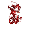 6fcaC  6fccC 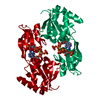 6fctC 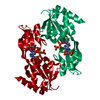 6fcwC  6fcyC  6fttC  6fu2C  6fu7C  6fuaC  5m8hS S: Starting model for refinement C: citing same article ( |
|---|---|
| Similar structure data |


-Links
 PDBj
PDBj


- Assembly
| Deposited unit | 
| ||||||||
|---|---|---|---|---|---|---|---|---|---|
| 1 | 
| ||||||||
| Unit cell |
| ||||||||
| Components on special symmetry positions |
|
-Components
| #1: Protein | / ATP-PRTase Mass: 25240.965 Da / Num. of mol.: 1 Source method: isolated from a genetically manipulated source Details: Residues from N- and C-termini are missing from coordinate sequence due to flexibility - electron density missing. Initial glycine remains after affinity-tag cleavage. Source: (gene. exp.) Psychrobacter arcticus (strain DSM 17307 / 273-4) (bacteria)Strain: DSM 17307 / 273-4 / Gene: hisG, Psyc_1903 / Production host: Escherichia coli BL21(DE3) (bacteria) / Strain (production host): C43 / References: UniProt: Q4FQF7, ATP phosphoribosyltransferase |
|---|---|
| #2: Chemical | ChemComp-AMP / Adenosine monophosphate  Mass: 347.221 Da / Num. of mol.: 1 / Source method: obtained synthetically / Formula: C10H14N5O7P / Feature type: SUBJECT OF INVESTIGATION / Comment: AMP*YM Mass: 347.221 Da / Num. of mol.: 1 / Source method: obtained synthetically / Formula: C10H14N5O7P / Feature type: SUBJECT OF INVESTIGATION / Comment: AMP*YM |
| #3: Water | ChemComp-HOH / Water Mass: 18.015 Da / Num. of mol.: 86 / Source method: isolated from a natural source / Formula: H2O Mass: 18.015 Da / Num. of mol.: 86 / Source method: isolated from a natural source / Formula: H2O |
-Experimental details
-Experiment
| Experiment | Method: X-RAY DIFFRACTION / Number of used crystals: 1 |
|---|
- Sample preparation
| Crystal | Density Matthews: 2.12 Å3/Da / Density % sol: 42.02 % |
|---|---|
| Crystal grow | Temperature: 277 K / Method: vapor diffusion / pH: 6.5 Details: 10mg/ml protein in 20mM Tris HCl pH8.0, 50mM KCl, 10mM MgCl2, 2mM DTT mixed 1:1 with 32% PEG 3350, 0.1M MOPS pH6.5, 0.1M K/Na tartrate |
-Data collection
| Diffraction | Mean temperature: 100 K |
|---|---|
| Diffraction source | Source: ROTATING ANODE / Type: RIGAKU MICROMAX-007 HF / Wavelength: 1.5418 Å |
| Detector | Type: RIGAKU SATURN 944+ / Detector: CCD / Date: Jun 6, 2017 / Details: mirrors |
| Radiation | Protocol: SINGLE WAVELENGTH / Monochromatic (M) / Laue (L): M / Scattering type: x-ray |
| Radiation wavelength | Wavelength: 1.5418 Å / Relative weight: 1 |
| Reflection | Resolution: 2.2→26.61 Å / Num. obs: 10299 / % possible obs: 93 % / Redundancy: 3.6 % / CC1/2: 0.981 / Rmerge(I) obs: 0.168 / Rpim(I) all: 0.105 / Rrim(I) all: 0.199 / Net I/σ(I): 5.8 / Num. measured all: 37047 / Scaling rejects: 9 |
| Reflection shell | Resolution: 2.2→2.27 Å / Redundancy: 3.6 % / Rmerge(I) obs: 0.549 / Num. unique obs: 827 / CC1/2: 0.706 / Rpim(I) all: 0.336 / Rrim(I) all: 0.645 / % possible all: 87.9 |
-Phasing
| Phasing | Method: molecular replacement |
|---|
- Processing
| Software |
| ||||||||||||||||||||||||||||||||||||||||||||||||||||||||||||
|---|---|---|---|---|---|---|---|---|---|---|---|---|---|---|---|---|---|---|---|---|---|---|---|---|---|---|---|---|---|---|---|---|---|---|---|---|---|---|---|---|---|---|---|---|---|---|---|---|---|---|---|---|---|---|---|---|---|---|---|---|---|
| Refinement | Method to determine structure: MOLECULAR REPLACEMENT Starting model: 5M8H chain E Resolution: 2.2→26.61 Å / Cor.coef. Fo:Fc: 0.947 / Cor.coef. Fo:Fc free: 0.911 / SU B: 7.367 / SU ML: 0.177 / Cross valid method: THROUGHOUT / σ(F): 0 / ESU R: 0.325 / ESU R Free: 0.223 Details: HYDROGENS HAVE BEEN ADDED IN THE RIDING POSITIONS U VALUES : REFINED INDIVIDUALLY
| ||||||||||||||||||||||||||||||||||||||||||||||||||||||||||||
| Solvent computation | Ion probe radii: 0.7 Å / Shrinkage radii: 0.7 Å / VDW probe radii: 1 Å | ||||||||||||||||||||||||||||||||||||||||||||||||||||||||||||
| Displacement parameters | Biso max: 64.29 Å2 / Biso mean: 25.922 Å2 / Biso min: 14.64 Å2
| ||||||||||||||||||||||||||||||||||||||||||||||||||||||||||||
| Refinement step | Cycle: final / Resolution: 2.2→26.61 Å
| ||||||||||||||||||||||||||||||||||||||||||||||||||||||||||||
| Refine LS restraints |
| ||||||||||||||||||||||||||||||||||||||||||||||||||||||||||||
| LS refinement shell | Resolution: 2.2→2.257 Å / Rfactor Rfree error: 0 / Total num. of bins used: 20
|
