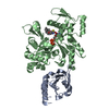
[English] 日本語
Yorodumi
- PDB-5zza: OdinProfilin/Rabbit Actin Complex -

+
Open data


ID or keywords:

Loading...

-
Basic information

Entry
Database: PDB / ID: 5zza
TitleOdinProfilin/Rabbit Actin Complex
Components
  • Actin, alpha skeletal muscle
  • Uncharacterized protein
KeywordsSTRUCTURAL PROTEIN / Actin / Archaea / Odin
Function / homology
Function and homology information


cytoskeletal motor activator activity / tropomyosin binding / myosin heavy chain binding / mesenchyme migration / troponin I binding / actin filament bundle / filamentous actin / actin filament bundle assembly / skeletal muscle thin filament assembly / striated muscle thin filament ...cytoskeletal motor activator activity / tropomyosin binding / myosin heavy chain binding / mesenchyme migration / troponin I binding / actin filament bundle / filamentous actin / actin filament bundle assembly / skeletal muscle thin filament assembly / striated muscle thin filament / skeletal muscle myofibril / actin monomer binding / skeletal muscle fiber development / stress fiber / titin binding / actin filament polymerization / filopodium / actin filament / Hydrolases; Acting on acid anhydrides; Acting on acid anhydrides to facilitate cellular and subcellular movement / calcium-dependent protein binding / lamellipodium / cell body / actin binding / cytoskeleton / hydrolase activity / protein domain specific binding / calcium ion binding / positive regulation of gene expression / magnesium ion binding / ATP binding / identical protein binding / cytoplasm
Similarity search - Function
Actin; Chain A, domain 2 / Actin; Chain A, domain 2 / Profilin superfamily / ATPase, substrate binding domain, subdomain 4 / Actin; Chain A, domain 4 / ATPase, nucleotide binding domain / Actins signature 1. / Actin, conserved site / Actins signature 2. / Actin/actin-like conserved site ...Actin; Chain A, domain 2 / Actin; Chain A, domain 2 / Profilin superfamily / ATPase, substrate binding domain, subdomain 4 / Actin; Chain A, domain 4 / ATPase, nucleotide binding domain / Actins signature 1. / Actin, conserved site / Actins signature 2. / Actin/actin-like conserved site / Actins and actin-related proteins signature. / Actin / Actin family / Actin / ATPase, nucleotide binding domain / Nucleotidyltransferase; domain 5 / Roll / Alpha-Beta Complex / 2-Layer Sandwich / Mainly Beta / Alpha Beta
Similarity search - Domain/homology
ADENOSINE-5'-TRIPHOSPHATE / LATRUNCULIN B / Odin profilin / Actin, alpha skeletal muscle
Similarity search - Component
Biological speciesCandidatus Odinarchaeota archaeon LCB_4 (archaea)
Oryctolagus cuniculus (rabbit)
MethodX-RAY DIFFRACTION / SYNCHROTRON / MOLECULAR REPLACEMENT / Resolution: 1.53 Å
AuthorsRobinson, R.C. / Akil, C.
CitationJournal: Nature / Year: 2018
Title: Genomes of Asgard archaea encode profilins that regulate actin.
Authors: Akil, C. / Robinson, R.C.
History
DepositionMay 31, 2018Deposition site: PDBJ / Processing site: PDBJ
Revision 1.0Oct 10, 2018Provider: repository / Type: Initial release
Revision 1.1Oct 17, 2018Group: Data collection / Database references / Structure summary
Category: citation / citation_author / entity
Item: _citation.pdbx_database_id_PubMed / _citation.title / _entity.formula_weight
Revision 1.2Oct 31, 2018Group: Data collection / Database references / Category: citation / citation_author / diffrn_detector
Item: _citation.journal_volume / _citation.page_first ..._citation.journal_volume / _citation.page_first / _citation.page_last / _diffrn_detector.detector / _diffrn_detector.type
Revision 1.3Dec 5, 2018Group: Data collection / Database references / Category: pdbx_related_exp_data_set
Revision 1.4Nov 22, 2023Group: Data collection / Database references ...Data collection / Database references / Derived calculations / Refinement description
Category: chem_comp_atom / chem_comp_bond ...chem_comp_atom / chem_comp_bond / database_2 / pdbx_initial_refinement_model / pdbx_struct_conn_angle / refine_hist / struct_conn
Item: _database_2.pdbx_DOI / _database_2.pdbx_database_accession ..._database_2.pdbx_DOI / _database_2.pdbx_database_accession / _pdbx_struct_conn_angle.ptnr1_auth_seq_id / _pdbx_struct_conn_angle.ptnr3_auth_seq_id / _pdbx_struct_conn_angle.value / _refine_hist.d_res_low / _struct_conn.pdbx_dist_value / _struct_conn.ptnr2_auth_seq_id

-
Structure visualization

Structure viewerMolecule:
MolmilJmol/JSmol

Downloads & links

-
Assembly

Deposited unit
P: Uncharacterized protein
A: Actin, alpha skeletal muscle
hetero molecules


Theoretical massNumber of molelcules
Total (without water)55,7846
Polymers54,4462
Non-polymers1,3384
Water13,673759
1


  • Idetical with deposited unit
  • defined by author&software
TypeNameSymmetry operationNumber
identity operation1_555x,y,z1
Buried area3420 Å2
ΔGint-11 kcal/mol
Surface area20800 Å2
MethodPISA
Unit cell
Length a, b, c (Å)54.079, 70.669, 77.717
Angle α, β, γ (deg.)90.00, 101.13, 90.00
Int Tables number4
Space group name H-MP1211

-
Components

-
Protein , 2 types, 2 molecules PA

#1: Protein Uncharacterized protein / OdinProfilin


Mass: 12814.657 Da / Num. of mol.: 1
Source method: isolated from a genetically manipulated source
Source: (gene. exp.) Candidatus Odinarchaeota archaeon LCB_4 (archaea)
Gene: OdinLCB4_14170 / Production host: Escherichia coli (E. coli) / References: UniProt: A0A1Q9N7W7
#2: Protein Actin, alpha skeletal muscle / / Alpha-actin-1


Mass: 41631.430 Da / Num. of mol.: 1 / Source method: isolated from a natural source / Source: (natural) Oryctolagus cuniculus (rabbit) / References: UniProt: P68135

-
Non-polymers , 4 types, 763 molecules

#3: Chemical ChemComp-LAB / LATRUNCULIN B / Latrunculin


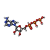
Mass: 395.513 Da / Num. of mol.: 2 / Source method: obtained synthetically / Formula: C20H29NO5S / Comment: toxin*YM
#4: Chemical ChemComp-ATP / ADENOSINE-5'-TRIPHOSPHATE / Adenosine triphosphate


Mass: 507.181 Da / Num. of mol.: 1 / Source method: obtained synthetically / Formula: C10H16N5O13P3 / Comment: ATP, energy-carrying molecule*YM
#5: Chemical ChemComp-CA / CALCIUM ION


Mass: 40.078 Da / Num. of mol.: 1 / Source method: obtained synthetically / Formula: Ca
#6: Water ChemComp-HOH / water / Water


Mass: 18.015 Da / Num. of mol.: 759 / Source method: isolated from a natural source / Formula: H2O

-
Experimental details

-
Experiment

ExperimentMethod: X-RAY DIFFRACTION / Number of used crystals: 1

-
Sample preparation

CrystalDensity Matthews: 2.68 Å3/Da / Density % sol: 54.18 %
Crystal growTemperature: 291 K / Method: vapor diffusion, sitting drop / pH: 5 / Details: 100 mM Citrate, 20% PEG6000

-
Data collection

DiffractionMean temperature: 100 K
Diffraction sourceSource: SYNCHROTRON / Site: Australian Synchrotron / Beamline: MX2 / Wavelength: 1 Å
DetectorType: DECTRIS EIGER X 16M / Detector: PIXEL / Date: Dec 7, 2017
RadiationProtocol: SINGLE WAVELENGTH / Monochromatic (M) / Laue (L): M / Scattering type: x-ray
Radiation wavelengthWavelength: 1 Å / Relative weight: 1
ReflectionResolution: 1.53→39.8 Å / Num. obs: 85118 / % possible obs: 98.7 % / Redundancy: 5.2 % / CC1/2: 0.999 / Rmerge(I) obs: 0.051 / Rpim(I) all: 0.024 / Rrim(I) all: 0.057 / Net I/av σ(I): 12.5 / Net I/σ(I): 12.5
Reflection shellResolution: 1.53→1.56 Å / Redundancy: 4.9 % / Rmerge(I) obs: 0.314 / Mean I/σ(I) obs: 2.3 / Num. unique obs: 1968 / CC1/2: 0.941 / Rpim(I) all: 0.152 / Rrim(I) all: 0.35 / % possible all: 81.7

-
Processing

Software
NameVersionClassification
REFMAC5.8.0189refinement
ADDREFdata reduction
Aimlessdata scaling
PHASERphasing
RefinementMethod to determine structure: MOLECULAR REPLACEMENT
Starting model: 5YEE
Resolution: 1.53→39.779 Å / Cor.coef. Fo:Fc: 0.974 / Cor.coef. Fo:Fc free: 0.965 / SU B: 2.722 / SU ML: 0.045 / Cross valid method: THROUGHOUT / ESU R: 0.084 / ESU R Free: 0.068 / Stereochemistry target values: MAXIMUM LIKELIHOOD / Details: HYDROGENS HAVE BEEN ADDED IN THE RIDING POSITIONS
RfactorNum. reflection% reflectionSelection details
Rfree0.17876 4265 5 %RANDOM
Rwork0.15041 ---
obs0.15182 80852 98.66 %-
Solvent computationIon probe radii: 0.8 Å / Shrinkage radii: 0.8 Å / VDW probe radii: 1.2 Å / Solvent model: MASK
Displacement parametersBiso mean: 22.246 Å2
Baniso -1Baniso -2Baniso -3
1--0.02 Å20 Å20.01 Å2
2--0.02 Å20 Å2
3---0 Å2
Refinement stepCycle: 1 / Resolution: 1.53→39.779 Å
ProteinNucleic acidLigandSolventTotal
Num. atoms3807 0 86 759 4652
Refine LS restraints
Refine-IDTypeDev idealDev ideal targetNumber
X-RAY DIFFRACTIONr_bond_refined_d0.0060.0194154
X-RAY DIFFRACTIONr_bond_other_d0.0020.023906
X-RAY DIFFRACTIONr_angle_refined_deg1.2981.9935675
X-RAY DIFFRACTIONr_angle_other_deg0.89939115
X-RAY DIFFRACTIONr_dihedral_angle_1_deg5.2725545
X-RAY DIFFRACTIONr_dihedral_angle_2_deg33.75624.128172
X-RAY DIFFRACTIONr_dihedral_angle_3_deg11.42515734
X-RAY DIFFRACTIONr_dihedral_angle_4_deg16.8751526
X-RAY DIFFRACTIONr_chiral_restr0.0710.2638
X-RAY DIFFRACTIONr_gen_planes_refined0.0040.0214630
X-RAY DIFFRACTIONr_gen_planes_other0.0030.02820
X-RAY DIFFRACTIONr_nbd_refined
X-RAY DIFFRACTIONr_nbd_other
X-RAY DIFFRACTIONr_nbtor_refined
X-RAY DIFFRACTIONr_nbtor_other
X-RAY DIFFRACTIONr_xyhbond_nbd_refined
X-RAY DIFFRACTIONr_xyhbond_nbd_other
X-RAY DIFFRACTIONr_metal_ion_refined
X-RAY DIFFRACTIONr_metal_ion_other
X-RAY DIFFRACTIONr_symmetry_vdw_refined
X-RAY DIFFRACTIONr_symmetry_vdw_other
X-RAY DIFFRACTIONr_symmetry_hbond_refined
X-RAY DIFFRACTIONr_symmetry_hbond_other
X-RAY DIFFRACTIONr_symmetry_metal_ion_refined
X-RAY DIFFRACTIONr_symmetry_metal_ion_other
X-RAY DIFFRACTIONr_mcbond_it0.7422.0822034
X-RAY DIFFRACTIONr_mcbond_other0.7422.0822033
X-RAY DIFFRACTIONr_mcangle_it0.8813.1192558
X-RAY DIFFRACTIONr_mcangle_other0.8813.1192559
X-RAY DIFFRACTIONr_scbond_it0.6982.1812120
X-RAY DIFFRACTIONr_scbond_other0.6982.182118
X-RAY DIFFRACTIONr_scangle_it
X-RAY DIFFRACTIONr_scangle_other0.7413.2183093
X-RAY DIFFRACTIONr_long_range_B_refined2.20826.8645044
X-RAY DIFFRACTIONr_long_range_B_other1.50925.0134721
X-RAY DIFFRACTIONr_rigid_bond_restr0.79138060
X-RAY DIFFRACTIONr_sphericity_free15.7625389
X-RAY DIFFRACTIONr_sphericity_bonded3.82958322
LS refinement shellResolution: 1.532→1.572 Å / Total num. of bins used: 20
RfactorNum. reflection% reflection
Rfree0.246 266 -
Rwork0.202 5273 -
obs--87.59 %

+
About Yorodumi

-
News

-
Feb 9, 2022. New format data for meta-information of EMDB entries

New format data for meta-information of EMDB entries

  • Version 3 of the EMDB header file is now the official format.
  • The previous official version 1.9 will be removed from the archive.

Related info.:EMDB header

External links:wwPDB to switch to version 3 of the EMDB data model

-
Aug 12, 2020. Covid-19 info

Covid-19 info

URL: https://pdbj.org/emnavi/covid19.php

New page: Covid-19 featured information page in EM Navigator.

Related info.:Covid-19 info / Mar 5, 2020. Novel coronavirus structure data

+
Mar 5, 2020. Novel coronavirus structure data

Novel coronavirus structure data

Related info.:Yorodumi Speices / Aug 12, 2020. Covid-19 info

External links:COVID-19 featured content - PDBj / Molecule of the Month (242):Coronavirus Proteases

+
Jan 31, 2019. EMDB accession codes are about to change! (news from PDBe EMDB page)

EMDB accession codes are about to change! (news from PDBe EMDB page)

  • The allocation of 4 digits for EMDB accession codes will soon come to an end. Whilst these codes will remain in use, new EMDB accession codes will include an additional digit and will expand incrementally as the available range of codes is exhausted. The current 4-digit format prefixed with “EMD-” (i.e. EMD-XXXX) will advance to a 5-digit format (i.e. EMD-XXXXX), and so on. It is currently estimated that the 4-digit codes will be depleted around Spring 2019, at which point the 5-digit format will come into force.
  • The EM Navigator/Yorodumi systems omit the EMD- prefix.

Related info.:Q: What is EMD? / ID/Accession-code notation in Yorodumi/EM Navigator

External links:EMDB Accession Codes are Changing Soon! / Contact to PDBj

+
Jul 12, 2017. Major update of PDB

Major update of PDB

  • wwPDB released updated PDB data conforming to the new PDBx/mmCIF dictionary.
  • This is a major update changing the version number from 4 to 5, and with Remediation, in which all the entries are updated.
  • In this update, many items about electron microscopy experimental information are reorganized (e.g. em_software).
  • Now, EM Navigator and Yorodumi are based on the updated data.

External links:wwPDB Remediation / Enriched Model Files Conforming to OneDep Data Standards Now Available in the PDB FTP Archive

-
Yorodumi

Thousand views of thousand structures

  • Yorodumi is a browser for structure data from EMDB, PDB, SASBDB, etc.
  • This page is also the successor to EM Navigator detail page, and also detail information page/front-end page for Omokage search.
  • The word "yorodu" (or yorozu) is an old Japanese word meaning "ten thousand". "mi" (miru) is to see.

Related info.:EMDB / PDB / SASBDB / Comparison of 3 databanks / Yorodumi Search / Aug 31, 2016. New EM Navigator & Yorodumi / Yorodumi Papers / Jmol/JSmol / Function and homology information / Changes in new EM Navigator and Yorodumi

Read more