 Movie
Movie Controller


+ Open data

- Basic information
| Entry | Database: PDB / ID: 5z9l | ||||||
|---|---|---|---|---|---|---|---|
| Title | Bacterial GyrB ATPase domain in complex with a chemical fragment | ||||||
 Components Components | DNA gyrase subunit B | ||||||
 Keywords Keywords | ISOMERASE / DNA Topoisomerase / Antibacterial / Drug Target / Fragment-base Lead Discovery | ||||||
| Function / homology |  Function and homology information Function and homology informationDNA topoisomerase type II (double strand cut, ATP-hydrolyzing) complex / DNA negative supercoiling activity / DNA topoisomerase type II (double strand cut, ATP-hydrolyzing) activity / DNA topoisomerase (ATP-hydrolysing) / DNA topological change / ATP-dependent activity, acting on DNA / DNA-templated DNA replication / chromosome / response to xenobiotic stimulus / response to antibiotic ...DNA topoisomerase type II (double strand cut, ATP-hydrolyzing) complex / DNA negative supercoiling activity / DNA topoisomerase type II (double strand cut, ATP-hydrolyzing) activity / DNA topoisomerase (ATP-hydrolysing) / DNA topological change / ATP-dependent activity, acting on DNA / DNA-templated DNA replication / chromosome / response to xenobiotic stimulus / response to antibiotic / DNA-templated transcription / DNA binding / ATP binding / metal ion binding / cytosol / cytoplasmSimilarity search - Function | ||||||
| Biological species |  Escherichia coli (E. coli) Escherichia coli (E. coli) | ||||||
| Method | X-RAY DIFFRACTION / SYNCHROTRON / MOLECULAR REPLACEMENT / Resolution: 1.6 Å | ||||||
 Authors Authors | Huang, X. / Zhou, H. | ||||||
| Funding support |  China, 1items China, 1items
| ||||||
 Citation Citation | Journal: Medchemcomm / Year: 2018 Title: Identification of an auxiliary druggable pocket in the DNA gyrase ATPase domain using fragment probes Authors: Huang, X. / Guo, J. / Liu, Q. / Gu, Q. / Xu, J. / Zhou, H. | ||||||
| History |
|
- Structure visualization
| Structure viewer | Molecule: MolmilJmol/JSmol |
|---|
- Downloads & links
-Download
| PDBx/mmCIF format | 5z9l.cif.gz | 155.7 KB | Display | PDBx/mmCIF format |
|---|---|---|---|---|
| PDB format | pdb5z9l.ent.gz | 122 KB | Display | PDB format |
| PDBx/mmJSON format | 5z9l.json.gz | Tree view | PDBx/mmJSON format | |
| Others |  Other downloads Other downloads |
-Validation report
| Arichive directory | https://data.pdbj.org/pub/pdb/validation_reports/z9/5z9lftp://data.pdbj.org/pub/pdb/validation_reports/z9/5z9l | HTTPS FTP |
|---|
-Related structure data
| Related structure data | 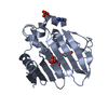 5z4hC  5z4oC  5z9bC  5z9eC  5z9fC  5z9mC  5z9nC  5z9pC  5z9qC 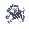 4duhS S: Starting model for refinement C: citing same article ( |
|---|---|
| Similar structure data |


-Links
 PDBj
PDBj


- Assembly
| Deposited unit | 
| ||||||||
|---|---|---|---|---|---|---|---|---|---|
| 1 | 
| ||||||||
| 2 | 
| ||||||||
| Unit cell |
|
-Components
| #1: Protein | / Type IIA topoisomerase subunit GyrB Mass: 22705.473 Da / Num. of mol.: 2 / Fragment: UNP residues 15-221 Source method: isolated from a genetically manipulated source Source: (gene. exp.) Escherichia coli (strain K12) (bacteria)Strain: K12 Gene: gyrB, acrB, cou, himB, hisU, nalC, parA, pcbA, b3699, JW5625 Production host: Escherichia coli (E. coli) / References: UniProt: P0AES6, EC: 5.99.1.3#2: Chemical | ChemComp-PO4 / Phosphate  Mass: 94.971 Da / Num. of mol.: 4 / Source method: obtained synthetically / Formula: PO4 Mass: 94.971 Da / Num. of mol.: 4 / Source method: obtained synthetically / Formula: PO4#3: Chemical |   Mass: 137.111 Da / Num. of mol.: 2 / Source method: obtained synthetically / Formula: C7H4FNO / Feature type: SUBJECT OF INVESTIGATION Mass: 137.111 Da / Num. of mol.: 2 / Source method: obtained synthetically / Formula: C7H4FNO / Feature type: SUBJECT OF INVESTIGATION#4: Chemical | ChemComp-AX7 / |   Mass: 133.151 Da / Num. of mol.: 1 / Source method: obtained synthetically / Formula: C7H7N3 Mass: 133.151 Da / Num. of mol.: 1 / Source method: obtained synthetically / Formula: C7H7N3#5: Water | ChemComp-HOH / | Water Mass: 18.015 Da / Num. of mol.: 160 / Source method: isolated from a natural source / Formula: H2O Mass: 18.015 Da / Num. of mol.: 160 / Source method: isolated from a natural source / Formula: H2O |
|---|
-Experimental details
-Experiment
| Experiment | Method: X-RAY DIFFRACTION / Number of used crystals: 1 |
|---|
- Sample preparation
| Crystal | Density Matthews: 2.37 Å3/Da / Density % sol: 48.13 % |
|---|---|
| Crystal grow | Temperature: 281 K / Method: vapor diffusion, sitting drop Details: 0.1M Tris-HCl pH 7.5, 2.20M (NH4)2HPO4, 10mM 2-aminobenzimidazole |
-Data collection
| Diffraction | Mean temperature: 100 K |
|---|---|
| Diffraction source | Source: SYNCHROTRON / Site: SSRF / Beamline: BL17U1 / Wavelength: 0.979 Å |
| Detector | Type: ADSC QUANTUM 315r / Detector: CCD / Date: May 2, 2017 |
| Radiation | Protocol: SINGLE WAVELENGTH / Monochromatic (M) / Laue (L): M / Scattering type: x-ray |
| Radiation wavelength | Wavelength: 0.979 Å / Relative weight: 1 |
| Reflection | Resolution: 1.6→57.22 Å / Num. obs: 55927 / % possible obs: 96.9 % / Redundancy: 5.2 % / Rmerge(I) obs: 0.071 / Net I/σ(I): 10.8 |
| Reflection shell | Resolution: 1.6→1.68 Å / Redundancy: 3.1 % / Rmerge(I) obs: 0.379 / Mean I/σ(I) obs: 2.7 / Num. unique obs: 7422 / % possible all: 89.6 |
- Processing
| Software |
| ||||||||||||||||||||||||||||||||||||||||||||||||||||||||||||||||||||||||||||||||||||||||||||||||||||||||||||||||||||||||||||||||||||||||||||||||||||||||||||||||||||||||||||||||||||||
|---|---|---|---|---|---|---|---|---|---|---|---|---|---|---|---|---|---|---|---|---|---|---|---|---|---|---|---|---|---|---|---|---|---|---|---|---|---|---|---|---|---|---|---|---|---|---|---|---|---|---|---|---|---|---|---|---|---|---|---|---|---|---|---|---|---|---|---|---|---|---|---|---|---|---|---|---|---|---|---|---|---|---|---|---|---|---|---|---|---|---|---|---|---|---|---|---|---|---|---|---|---|---|---|---|---|---|---|---|---|---|---|---|---|---|---|---|---|---|---|---|---|---|---|---|---|---|---|---|---|---|---|---|---|---|---|---|---|---|---|---|---|---|---|---|---|---|---|---|---|---|---|---|---|---|---|---|---|---|---|---|---|---|---|---|---|---|---|---|---|---|---|---|---|---|---|---|---|---|---|---|---|---|---|
| Refinement | Method to determine structure: MOLECULAR REPLACEMENT Starting model: 4DUH Resolution: 1.6→57.21 Å / Cor.coef. Fo:Fc: 0.928 / Cor.coef. Fo:Fc free: 0.924 / SU B: 4.004 / SU ML: 0.072 / Cross valid method: THROUGHOUT / ESU R: 0.103 / ESU R Free: 0.096 / Details: HYDROGENS HAVE BEEN ADDED IN THE RIDING POSITIONS
| ||||||||||||||||||||||||||||||||||||||||||||||||||||||||||||||||||||||||||||||||||||||||||||||||||||||||||||||||||||||||||||||||||||||||||||||||||||||||||||||||||||||||||||||||||||||
| Solvent computation | Ion probe radii: 0.8 Å / Shrinkage radii: 0.8 Å / VDW probe radii: 1.2 Å | ||||||||||||||||||||||||||||||||||||||||||||||||||||||||||||||||||||||||||||||||||||||||||||||||||||||||||||||||||||||||||||||||||||||||||||||||||||||||||||||||||||||||||||||||||||||
| Displacement parameters | Biso mean: 23.039 Å2
| ||||||||||||||||||||||||||||||||||||||||||||||||||||||||||||||||||||||||||||||||||||||||||||||||||||||||||||||||||||||||||||||||||||||||||||||||||||||||||||||||||||||||||||||||||||||
| Refinement step | Cycle: 1 / Resolution: 1.6→57.21 Å
| ||||||||||||||||||||||||||||||||||||||||||||||||||||||||||||||||||||||||||||||||||||||||||||||||||||||||||||||||||||||||||||||||||||||||||||||||||||||||||||||||||||||||||||||||||||||
| Refine LS restraints |
|