| Entry | Database: PDB / ID: 5yt0
|
|---|
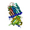
| Title | Crystal structure of the complex of archaeal ribosomal stalk protein aP1 and archaeal translation initiation factor aIF5B |
|---|
 Components Components | - Archaeal ribosomal stalk protein aP1
- Probable translation initiation factor IF-2
|
|---|
 Keywords Keywords |  TRANSLATION / translation initiation factor / ribosomal stalk protein / complex TRANSLATION / translation initiation factor / ribosomal stalk protein / complex |
|---|
| Function / homology |  Function and homology information Function and homology information
translational elongation / translation initiation factor activity / ribosome / structural constituent of ribosome / ribonucleoprotein complex / GTPase activity / GTP bindingSimilarity search - Function Ribosomal protein L12, archaea / Translation initiation factor aIF-2, archaea / Ribosomal protein L12/P1/P2 family / Ribosomal protein P1/P2, N-terminal domain / Elongation factor Tu-type domain / Elongation factor Tu domain 4 / Translation initiation factor IF- 2, domain 3 / Translation-initiation factor 2 / Translation initiation factor IF- 2 / Translation initiation factor IF-2, domain 3 superfamily ...Ribosomal protein L12, archaea / Translation initiation factor aIF-2, archaea / Ribosomal protein L12/P1/P2 family / Ribosomal protein P1/P2, N-terminal domain / Elongation factor Tu-type domain / Elongation factor Tu domain 4 / Translation initiation factor IF- 2, domain 3 / Translation-initiation factor 2 / Translation initiation factor IF- 2 / Translation initiation factor IF-2, domain 3 superfamily / 60s Acidic ribosomal protein / Translation factors / Translation elongation factor EFTu-like, domain 2 / Elongation factor Tu domain 2 / Translational (tr)-type GTP-binding domain / Elongation factor Tu GTP binding domain / Translational (tr)-type guanine nucleotide-binding (G) domain profile. / Elongation Factor Tu (Ef-tu); domain 3 / Small GTP-binding protein domain / Translation protein, beta-barrel domain superfamily / P-loop containing nucleotide triphosphate hydrolases / Beta Barrel / P-loop containing nucleoside triphosphate hydrolase / Rossmann fold / 3-Layer(aba) Sandwich / Mainly Beta / Alpha BetaSimilarity search - Domain/homology |
|---|
| Biological species |    Aeropyrum pernix K1 (archaea) Aeropyrum pernix K1 (archaea) |
|---|
| Method | X-RAY DIFFRACTION / SYNCHROTRON / MOLECULAR REPLACEMENT / Resolution: 1.89 Å |
|---|
 Authors Authors | Murakami, R. / Singh, C.R. / Morris, J. / Tang, L. / Harmon, I. / Miyoshi, T. / Ito, K. / Asano, K. / Uchiumi, T. |
|---|
| Funding support |  Japan, 1items Japan, 1items | Organization | Grant number | Country |
|---|
| the Japan Society for the Promotion of Science | 15K06964 | Japan |
|
|---|
 Citation Citation | Journal: Mol. Cell. Biol. / Year: 2018
Title: The Interaction between the Ribosomal Stalk Proteins and Translation Initiation Factor 5B Promotes Translation Initiation
Authors: Murakami, R. / Singh, C.R. / Morris, J. / Tang, L. / Harmon, I. / Takasu, A. / Miyoshi, T. / Ito, K. / Asano, K. / Uchiumi, T. |
|---|
| History | | Deposition | Nov 16, 2017 | Deposition site: PDBJ / Processing site: PDBJ |
|---|
| Revision 1.0 | Jun 27, 2018 | Provider: repository / Type: Initial release |
|---|
| Revision 1.1 | Aug 15, 2018 | Group: Data collection / Database references / Category: citation / citation_author
Item: _citation.journal_volume / _citation.title / _citation_author.name |
|---|
| Revision 1.2 | Mar 27, 2024 | Group: Data collection / Database references / Category: chem_comp_atom / chem_comp_bond / database_2
Item: _database_2.pdbx_DOI / _database_2.pdbx_database_accession |
|---|
|
|---|
 Movie
Movie Controller

 Yorodumi
 Open data
 Basic information
 Structure visualization
 Downloads & links
 Other downloads


 PDBj
PDBj


 Assembly

 Type: RNA linking / Mass: 443.201 Da / Num. of mol.: 1 / Source method: obtained synthetically / Formula: C10H15N5O11P2 / Comment: GDP, energy-carrying molecule*YM
 Mass: 18.015 Da / Num. of mol.: 151 / Source method: isolated from a natural source / Formula: H2O
 Sample preparation
 Processing