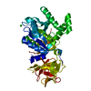
[English] 日本語
Yorodumi
- PDB-5vh6: 2.6 Angstrom Resolution Crystal Structure of N-terminal Fragment ... -

+
Open data


ID or keywords:

Loading...

-
Basic information

Entry
Database: PDB / ID: 5vh6
Title2.6 Angstrom Resolution Crystal Structure of N-terminal Fragment (residues 1-406) of Elongation Factor G from Bacillus subtilis.
ComponentsElongation factor GEF-G
KeywordsHYDROLASE / OXIDOREDUCTASE / Structural Genomics / Center for Structural Genomics of Infectious Diseases / CSGID / Elongation Factor G
Function / homology
Function and homology information


ribosome disassembly / translation elongation factor activity / GTPase activity / GTP binding / cytosol
Similarity search - Function
Translation elongation factor EFG/EF2 / Elongation factor G, domain III / EFG, domain V / Elongation Factor G, domain II / Elongation Factor G, domain III / Translation elongation factor EFG/EF2, domain IV / Elongation factor G, domain IV / Elongation factor G, domain IV / Elongation factor G C-terminus / Elongation factor EFG, domain V-like ...Translation elongation factor EFG/EF2 / Elongation factor G, domain III / EFG, domain V / Elongation Factor G, domain II / Elongation Factor G, domain III / Translation elongation factor EFG/EF2, domain IV / Elongation factor G, domain IV / Elongation factor G, domain IV / Elongation factor G C-terminus / Elongation factor EFG, domain V-like / Elongation factor G C-terminus / Translation factors / EF-G domain III/V-like / Tr-type G domain, conserved site / Translational (tr)-type guanine nucleotide-binding (G) domain signature. / Translation elongation factor EFTu-like, domain 2 / Elongation factor Tu domain 2 / Translational (tr)-type GTP-binding domain / Elongation factor Tu GTP binding domain / Translational (tr)-type guanine nucleotide-binding (G) domain profile. / Elongation Factor Tu (Ef-tu); domain 3 / Small GTP-binding protein domain / Translation protein, beta-barrel domain superfamily / Ribosomal protein S5 domain 2-type fold, subgroup / P-loop containing nucleotide triphosphate hydrolases / Ribosomal protein S5 domain 2-type fold / Beta Barrel / P-loop containing nucleoside triphosphate hydrolase / Rossmann fold / 3-Layer(aba) Sandwich / Mainly Beta / Alpha Beta
Similarity search - Domain/homology
Biological speciesBacillus subtilis (bacteria)
MethodX-RAY DIFFRACTION / SYNCHROTRON / MOLECULAR REPLACEMENT / Resolution: 2.61 Å
AuthorsMinasov, G. / Shuvalova, L. / Dubrovska, I. / Kiryukhina, O. / Grimshaw, S. / Kwon, K. / Anderson, W.F. / Center for Structural Genomics of Infectious Diseases (CSGID)
CitationJournal: To Be Published
Title: 2.6 Angstrom Resolution Crystal Structure of N-terminal Fragment (residues 1-406) of Elongation Factor G from Bacillus subtilis.
Authors: Minasov, G. / Shuvalova, L. / Dubrovska, I. / Kiryukhina, O. / Grimshaw, S. / Kwon, K. / Anderson, W.F. / Center for Structural Genomics of Infectious Diseases (CSGID)
History
DepositionApr 12, 2017Deposition site: RCSB / Processing site: RCSB
Revision 1.0Apr 19, 2017Provider: repository / Type: Initial release
Revision 1.1Oct 4, 2023Group: Data collection / Database references / Refinement description
Category: chem_comp_atom / chem_comp_bond ...chem_comp_atom / chem_comp_bond / database_2 / pdbx_initial_refinement_model
Item: _database_2.pdbx_DOI / _database_2.pdbx_database_accession

-
Structure visualization

Structure viewerMolecule:
MolmilJmol/JSmol

Downloads & links

-
Assembly

Deposited unit
A: Elongation factor G
hetero molecules


Theoretical massNumber of molelcules
Total (without water)45,6323
Polymers45,5611
Non-polymers712
Water1,13563
1


  • Idetical with deposited unit
  • defined by author&software
  • Evidence: homology, 5TY0
TypeNameSymmetry operationNumber
identity operation1_555x,y,z1
MethodPISA
Unit cell
Length a, b, c (Å)56.672, 56.672, 248.722
Angle α, β, γ (deg.)90.00, 90.00, 90.00
Int Tables number96
Space group name H-MP43212

-
Components

#1: Protein Elongation factor G / EF-G / EF-G / Vegetative protein 19 / VEG19


Mass: 45560.898 Da / Num. of mol.: 1 / Fragment: UNP residues 1-406
Source method: isolated from a genetically manipulated source
Source: (gene. exp.) Bacillus subtilis (strain 168) (bacteria)
Strain: 168 / Gene: fusA, fus, BSU01120 / Plasmid: pMCSG53 / Production host: Escherichia coli BL21(DE3) (bacteria) / References: UniProt: P80868
#2: Chemical ChemComp-CL / CHLORIDE ION / Chloride


Mass: 35.453 Da / Num. of mol.: 2 / Source method: obtained synthetically / Formula: Cl
#3: Water ChemComp-HOH / water / Water


Mass: 18.015 Da / Num. of mol.: 63 / Source method: isolated from a natural source / Formula: H2O

-
Experimental details

-
Experiment

ExperimentMethod: X-RAY DIFFRACTION / Number of used crystals: 1

-
Sample preparation

CrystalDensity Matthews: 2.2 Å3/Da / Density % sol: 43.4 %
Crystal growTemperature: 295 K / Method: vapor diffusion, sitting drop / pH: 6
Details: Protein: 8.0 mg/ml, 0.01M Tris HCl (pH 8.3); Screen: PACT (B7), 0.2M Sodium chloride, 0.1M MES pH (6.0), 20% (w/v) PEG 6000.

-
Data collection

DiffractionMean temperature: 100 K
Diffraction sourceSource: SYNCHROTRON / Site: APS / Beamline: 21-ID-F / Wavelength: 0.97872 Å
DetectorType: MARMOSAIC 300 mm CCD / Detector: CCD / Date: Mar 28, 2017 / Details: C(111)
RadiationMonochromator: Be / Protocol: SINGLE WAVELENGTH / Monochromatic (M) / Laue (L): M / Scattering type: x-ray
Radiation wavelengthWavelength: 0.97872 Å / Relative weight: 1
ReflectionResolution: 2.6→30 Å / Num. obs: 13280 / % possible obs: 99.8 % / Observed criterion σ(I): -3 / Redundancy: 6.8 % / Biso Wilson estimate: 59.2 Å2 / Rmerge(I) obs: 0.093 / Rpim(I) all: 0.038 / Rsym value: 0.093 / Χ2: 1.01 / Net I/σ(I): 18.5
Reflection shellResolution: 2.6→2.64 Å / Redundancy: 6.4 % / Rmerge(I) obs: 0.796 / Mean I/σ(I) obs: 2.2 / Num. unique obs: 638 / CC1/2: 0.867 / Rpim(I) all: 0.333 / Rsym value: 0.796 / Χ2: 1.003 / % possible all: 100

-
Processing

Software
NameVersionClassification
REFMAC5.8.0158refinement
HKL-3000data reduction
HKL-3000data scaling
MOLREPphasing
RefinementMethod to determine structure: MOLECULAR REPLACEMENT
Starting model: 2XEX
Resolution: 2.61→28.81 Å / Cor.coef. Fo:Fc: 0.947 / Cor.coef. Fo:Fc free: 0.92 / SU B: 32.689 / SU ML: 0.326 / Cross valid method: THROUGHOUT / ESU R: 1.395 / ESU R Free: 0.36 / Details: HYDROGENS HAVE BEEN ADDED IN THE RIDING POSITIONS
RfactorNum. reflection% reflectionSelection details
Rfree0.27732 661 5 %RANDOM
Rwork0.22542 ---
obs0.22811 12544 99.59 %-
Solvent computationIon probe radii: 0.8 Å / Shrinkage radii: 0.8 Å / VDW probe radii: 1.2 Å
Displacement parametersBiso mean: 75.252 Å2
Baniso -1Baniso -2Baniso -3
1-3.79 Å20 Å20 Å2
2--3.79 Å20 Å2
3----7.59 Å2
Refinement stepCycle: 1 / Resolution: 2.61→28.81 Å
ProteinNucleic acidLigandSolventTotal
Num. atoms2933 0 2 63 2998
Refine LS restraints
Refine-IDTypeDev idealDev ideal targetNumber
X-RAY DIFFRACTIONr_bond_refined_d0.0060.0192978
X-RAY DIFFRACTIONr_bond_other_d0.0010.022791
X-RAY DIFFRACTIONr_angle_refined_deg1.2381.9684033
X-RAY DIFFRACTIONr_angle_other_deg0.80936463
X-RAY DIFFRACTIONr_dihedral_angle_1_deg1.575374
X-RAY DIFFRACTIONr_dihedral_angle_2_deg17.04224.571140
X-RAY DIFFRACTIONr_dihedral_angle_3_deg7.10215522
X-RAY DIFFRACTIONr_dihedral_angle_4_deg9.0921521
X-RAY DIFFRACTIONr_chiral_restr0.0680.2467
X-RAY DIFFRACTIONr_gen_planes_refined0.0210.023327
X-RAY DIFFRACTIONr_gen_planes_other0.0180.02590
X-RAY DIFFRACTIONr_nbd_refined
X-RAY DIFFRACTIONr_nbd_other
X-RAY DIFFRACTIONr_nbtor_refined
X-RAY DIFFRACTIONr_nbtor_other
X-RAY DIFFRACTIONr_xyhbond_nbd_refined
X-RAY DIFFRACTIONr_xyhbond_nbd_other
X-RAY DIFFRACTIONr_metal_ion_refined
X-RAY DIFFRACTIONr_metal_ion_other
X-RAY DIFFRACTIONr_symmetry_vdw_refined
X-RAY DIFFRACTIONr_symmetry_vdw_other
X-RAY DIFFRACTIONr_symmetry_hbond_refined
X-RAY DIFFRACTIONr_symmetry_hbond_other
X-RAY DIFFRACTIONr_symmetry_metal_ion_refined
X-RAY DIFFRACTIONr_symmetry_metal_ion_other
X-RAY DIFFRACTIONr_mcbond_it1.8514.9621502
X-RAY DIFFRACTIONr_mcbond_other1.8444.9591501
X-RAY DIFFRACTIONr_mcangle_it3.3247.4291874
X-RAY DIFFRACTIONr_mcangle_other3.3247.4331875
X-RAY DIFFRACTIONr_scbond_it1.445.1581475
X-RAY DIFFRACTIONr_scbond_other1.4395.1621476
X-RAY DIFFRACTIONr_scangle_it
X-RAY DIFFRACTIONr_scangle_other2.6267.672160
X-RAY DIFFRACTIONr_long_range_B_refined5.61359.2653288
X-RAY DIFFRACTIONr_long_range_B_other5.61259.273285
X-RAY DIFFRACTIONr_rigid_bond_restr
X-RAY DIFFRACTIONr_sphericity_free
X-RAY DIFFRACTIONr_sphericity_bonded
LS refinement shellResolution: 2.607→2.674 Å / Total num. of bins used: 20
RfactorNum. reflection% reflection
Rfree0.298 48 -
Rwork0.354 889 -
obs--99.05 %
Refinement TLS params.

Method: refined / Refine-ID: X-RAY DIFFRACTION

IDL112)L122)L132)L222)L232)L332)S11 (Å °)S12 (Å °)S13 (Å °)S21 (Å °)S22 (Å °)S23 (Å °)S31 (Å °)S32 (Å °)S33 (Å °)T112)T122)T132)T222)T232)T332)Origin x (Å)Origin y (Å)Origin z (Å)
12.83740.4979-1.32963.72441.78127.44610.01120.0113-0.05160.3031-0.0381-0.0081.12270.23020.02690.26980.0342-0.00330.0476-0.00880.00330.6472-27.0531-15.3566
21.56151.63670.2723.07480.735110.26440.00330.0969-0.22870.52180.3879-0.66891.40181.3041-0.39120.50930.3638-0.18060.2994-0.14050.188412.0647-27.5826-2.9368
30.3060.035-0.67471.03110.96366.40010.0155-0.10630.16710.21940.2621-0.1490.63370.2539-0.27760.24930.0306-0.03730.1986-0.07230.17962.7113-16.5951-0.5174
43.95040.3267-1.53053.27340.42396.25580.10030.3086-0.1341-0.1147-0.01240.07330.0207-0.3481-0.08790.06510.0535-0.0320.1216-0.00660.04672.8728-24.7039-33.187
Refinement TLS group
IDRefine-IDRefine TLS-IDAuth asym-IDAuth seq-ID
1X-RAY DIFFRACTION1A0 - 95
2X-RAY DIFFRACTION2A96 - 157
3X-RAY DIFFRACTION3A158 - 300
4X-RAY DIFFRACTION4A301 - 400

+
About Yorodumi

-
News

-
Feb 9, 2022. New format data for meta-information of EMDB entries

New format data for meta-information of EMDB entries

  • Version 3 of the EMDB header file is now the official format.
  • The previous official version 1.9 will be removed from the archive.

Related info.:EMDB header

External links:wwPDB to switch to version 3 of the EMDB data model

-
Aug 12, 2020. Covid-19 info

Covid-19 info

URL: https://pdbj.org/emnavi/covid19.php

New page: Covid-19 featured information page in EM Navigator.

Related info.:Covid-19 info / Mar 5, 2020. Novel coronavirus structure data

+
Mar 5, 2020. Novel coronavirus structure data

Novel coronavirus structure data

Related info.:Yorodumi Speices / Aug 12, 2020. Covid-19 info

External links:COVID-19 featured content - PDBj / Molecule of the Month (242):Coronavirus Proteases

+
Jan 31, 2019. EMDB accession codes are about to change! (news from PDBe EMDB page)

EMDB accession codes are about to change! (news from PDBe EMDB page)

  • The allocation of 4 digits for EMDB accession codes will soon come to an end. Whilst these codes will remain in use, new EMDB accession codes will include an additional digit and will expand incrementally as the available range of codes is exhausted. The current 4-digit format prefixed with “EMD-” (i.e. EMD-XXXX) will advance to a 5-digit format (i.e. EMD-XXXXX), and so on. It is currently estimated that the 4-digit codes will be depleted around Spring 2019, at which point the 5-digit format will come into force.
  • The EM Navigator/Yorodumi systems omit the EMD- prefix.

Related info.:Q: What is EMD? / ID/Accession-code notation in Yorodumi/EM Navigator

External links:EMDB Accession Codes are Changing Soon! / Contact to PDBj

+
Jul 12, 2017. Major update of PDB

Major update of PDB

  • wwPDB released updated PDB data conforming to the new PDBx/mmCIF dictionary.
  • This is a major update changing the version number from 4 to 5, and with Remediation, in which all the entries are updated.
  • In this update, many items about electron microscopy experimental information are reorganized (e.g. em_software).
  • Now, EM Navigator and Yorodumi are based on the updated data.

External links:wwPDB Remediation / Enriched Model Files Conforming to OneDep Data Standards Now Available in the PDB FTP Archive

-
Yorodumi

Thousand views of thousand structures

  • Yorodumi is a browser for structure data from EMDB, PDB, SASBDB, etc.
  • This page is also the successor to EM Navigator detail page, and also detail information page/front-end page for Omokage search.
  • The word "yorodu" (or yorozu) is an old Japanese word meaning "ten thousand". "mi" (miru) is to see.

Related info.:EMDB / PDB / SASBDB / Comparison of 3 databanks / Yorodumi Search / Aug 31, 2016. New EM Navigator & Yorodumi / Yorodumi Papers / Jmol/JSmol / Function and homology information / Changes in new EM Navigator and Yorodumi

Read more