 Movie
Movie Controller


+ Open data

- Basic information
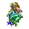
| Entry | Database: PDB / ID: 5v9j | ||||||
|---|---|---|---|---|---|---|---|
| Title | Crystal structure of catalytic domain of GLP with MS0105 | ||||||
 Components Components | Histone-lysine N-methyltransferase EHMT1 | ||||||
 Keywords Keywords |  TRANSFERASE / EHMT1 / methyltransferase / Structural Genomics / Structural Genomics Consortium / SGC TRANSFERASE / EHMT1 / methyltransferase / Structural Genomics / Structural Genomics Consortium / SGC | ||||||
| Function / homology |  Function and homology information Function and homology information[histone H3]-lysine9 N-methyltransferase / peptidyl-lysine monomethylation / histone H3K9 methyltransferase activity / histone H3K9me2 methyltransferase activity / peptidyl-lysine dimethylation / histone H3K27 methyltransferase activity / DNA methylation-dependent heterochromatin formation / protein-lysine N-methyltransferase activity / C2H2 zinc finger domain binding / Transcriptional Regulation by E2F6 ...[histone H3]-lysine9 N-methyltransferase / peptidyl-lysine monomethylation / histone H3K9 methyltransferase activity / histone H3K9me2 methyltransferase activity / peptidyl-lysine dimethylation / histone H3K27 methyltransferase activity / DNA methylation-dependent heterochromatin formation / protein-lysine N-methyltransferase activity / C2H2 zinc finger domain binding / Transcriptional Regulation by E2F6 / regulation of embryonic development / Transcriptional Regulation by VENTX / response to fungicide / Transferases; Transferring one-carbon groups; Methyltransferases / transcription corepressor binding / methyltransferase activity / Regulation of TP53 Activity through Methylation / PKMTs methylate histone lysines / p53 binding / chromatin organization / positive regulation of cold-induced thermogenesis / Senescence-Associated Secretory Phenotype (SASP) / nuclear body / negative regulation of DNA-templated transcription / chromatin / negative regulation of transcription by RNA polymerase II / zinc ion binding / nucleoplasm / nucleusSimilarity search - Function | ||||||
| Biological species |  Homo sapiens (human) Homo sapiens (human) | ||||||
| Method | X-RAY DIFFRACTION / SYNCHROTRON / FOURIER SYNTHESIS / Resolution: 1.74 Å | ||||||
 Authors Authors | Dong, A. / Zeng, H. / Liu, J. / Xiong, Y. / Babault, N. / Jin, J. / Tempel, W. / Bountra, C. / Arrowsmith, C.H. / Edwards, A.M. ...Dong, A. / Zeng, H. / Liu, J. / Xiong, Y. / Babault, N. / Jin, J. / Tempel, W. / Bountra, C. / Arrowsmith, C.H. / Edwards, A.M. / Wu, H. / Brown, P.J. / Structural Genomics Consortium (SGC) | ||||||
 Citation Citation | Journal: to be published Title: Crystal structure of catalytic domain of GLP with MS0105 Authors: Zeng, H. / Dong, A. / Liu, J. / Xiong, Y. / Babault, N. / Jin, J. / Tempel, W. / Bountra, C. / Arrowsmith, C.H. / Edwards, A.M. / Wu, H. / Brown, P.J. / Structural Genomics Consortium (SGC) | ||||||
| History |
|
- Structure visualization
| Structure viewer | Molecule: MolmilJmol/JSmol |
|---|
- Downloads & links
-Download
| PDBx/mmCIF format | 5v9j.cif.gz | 145.9 KB | Display | PDBx/mmCIF format |
|---|---|---|---|---|
| PDB format | pdb5v9j.ent.gz | 108.9 KB | Display | PDB format |
| PDBx/mmJSON format | 5v9j.json.gz | Tree view | PDBx/mmJSON format | |
| Others |  Other downloads Other downloads |
-Validation report
| Arichive directory | https://data.pdbj.org/pub/pdb/validation_reports/v9/5v9jftp://data.pdbj.org/pub/pdb/validation_reports/v9/5v9j | HTTPS FTP |
|---|
-Related structure data
| Related structure data |  5ttgS S: Starting model for refinement |
|---|---|
| Similar structure data |


-Links
 PDBj
PDBj


- Assembly
| Deposited unit | 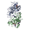
| ||||||||
|---|---|---|---|---|---|---|---|---|---|
| 1 |
| ||||||||
| Unit cell |
| ||||||||
| Details | THE AUTHORS STATE THAT THE BIOLOGICAL UNIT IS UNKNOWN. |
-Components
-Protein , 1 types, 2 molecules AB
| #1: Protein | Mass: 32974.344 Da / Num. of mol.: 2 / Fragment: residues 982-1266 Source method: isolated from a genetically manipulated source Source: (gene. exp.) Homo sapiens (human) / Gene: EHMT1, EUHMTASE1, GLP, KIAA1876, KMT1D / Plasmid: PET28-LIC / Production host:  Escherichia coli BL21 (bacteria) / Strain (production host): BL21-V2R-PRARE2 Escherichia coli BL21 (bacteria) / Strain (production host): BL21-V2R-PRARE2References: UniProt: Q9H9B1, Transferases; Transferring one-carbon groups; Methyltransferases, histone-lysine N-methyltransferase |
|---|
-Non-polymers , 7 types, 660 molecules 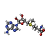


| #2: Chemical | S-Adenosyl methionine Mass: 398.437 Da / Num. of mol.: 2 / Source method: obtained synthetically / Formula: C15H22N6O5S Mass: 398.437 Da / Num. of mol.: 2 / Source method: obtained synthetically / Formula: C15H22N6O5S#3: Chemical |  Mass: 427.583 Da / Num. of mol.: 2 / Source method: obtained synthetically / Formula: C24H37N5O2 Mass: 427.583 Da / Num. of mol.: 2 / Source method: obtained synthetically / Formula: C24H37N5O2#4: Chemical | ChemComp-ZN /  Mass: 65.409 Da / Num. of mol.: 8 / Source method: obtained synthetically / Formula: Zn Mass: 65.409 Da / Num. of mol.: 8 / Source method: obtained synthetically / Formula: Zn#5: Chemical | Chloride Mass: 35.453 Da / Num. of mol.: 3 / Source method: obtained synthetically / Formula: Cl Mass: 35.453 Da / Num. of mol.: 3 / Source method: obtained synthetically / Formula: Cl#6: Chemical | ChemComp-UNX /  Num. of mol.: 13 / Source method: obtained synthetically Num. of mol.: 13 / Source method: obtained synthetically#7: Chemical | ChemComp-DMS / | Dimethyl sulfoxide Mass: 78.133 Da / Num. of mol.: 1 / Source method: obtained synthetically / Formula: C2H6OS / Comment: DMSO, precipitant*YM Mass: 78.133 Da / Num. of mol.: 1 / Source method: obtained synthetically / Formula: C2H6OS / Comment: DMSO, precipitant*YM#8: Water | ChemComp-HOH / | WaterMass: 18.015 Da / Num. of mol.: 631 / Source method: isolated from a natural source / Formula: H2O |
|---|
-Experimental details
-Experiment
| Experiment | Method: X-RAY DIFFRACTION / Number of used crystals: 1 |
|---|
- Sample preparation
| Crystal | Density Matthews: 2.79 Å3/Da / Density % sol: 55.86 % |
|---|---|
| Crystal grow | Temperature: 293 K / Method: vapor diffusion, sitting drop / pH: 5.6 / Details: 20% PEG 4000, 20% IProp, 0.1 M NaCitrate pH5.6 |
-Data collection
| Diffraction | Mean temperature: 100 K | ||||||||||||||||||||||||||||||||||||||||||||||||||||||||||||||||||||||||||||||||||||||||||||||||||||||||||||||||||||||||||||||
|---|---|---|---|---|---|---|---|---|---|---|---|---|---|---|---|---|---|---|---|---|---|---|---|---|---|---|---|---|---|---|---|---|---|---|---|---|---|---|---|---|---|---|---|---|---|---|---|---|---|---|---|---|---|---|---|---|---|---|---|---|---|---|---|---|---|---|---|---|---|---|---|---|---|---|---|---|---|---|---|---|---|---|---|---|---|---|---|---|---|---|---|---|---|---|---|---|---|---|---|---|---|---|---|---|---|---|---|---|---|---|---|---|---|---|---|---|---|---|---|---|---|---|---|---|---|---|---|
| Diffraction source | Source: SYNCHROTRON / Site: CLSI  / Beamline: 08ID-1 / Wavelength: 0.97949 Å / Beamline: 08ID-1 / Wavelength: 0.97949 Å | ||||||||||||||||||||||||||||||||||||||||||||||||||||||||||||||||||||||||||||||||||||||||||||||||||||||||||||||||||||||||||||||
| Detector | Type: RAYONIX MX-300 / Detector: CCD / Date: Sep 16, 2014 | ||||||||||||||||||||||||||||||||||||||||||||||||||||||||||||||||||||||||||||||||||||||||||||||||||||||||||||||||||||||||||||||
| Radiation | Protocol: SINGLE WAVELENGTH / Monochromatic (M) / Laue (L): M / Scattering type: x-ray | ||||||||||||||||||||||||||||||||||||||||||||||||||||||||||||||||||||||||||||||||||||||||||||||||||||||||||||||||||||||||||||||
| Radiation wavelength | Wavelength: 0.97949 Å / Relative weight: 1 | ||||||||||||||||||||||||||||||||||||||||||||||||||||||||||||||||||||||||||||||||||||||||||||||||||||||||||||||||||||||||||||||
| Reflection | Resolution: 1.74→50 Å / Num. obs: 76349 / % possible obs: 100 % / Redundancy: 8.1 % / Rmerge(I) obs: 0.123 / Χ2: 1.505 / Net I/av σ(I): 23.333 / Net I/σ(I): 7.4 | ||||||||||||||||||||||||||||||||||||||||||||||||||||||||||||||||||||||||||||||||||||||||||||||||||||||||||||||||||||||||||||||
| Reflection shell |
|
- Processing
| Software |
| |||||||||||||||||||||||||||||||||||||||||||||||||||||||||||||||||||||||||||
|---|---|---|---|---|---|---|---|---|---|---|---|---|---|---|---|---|---|---|---|---|---|---|---|---|---|---|---|---|---|---|---|---|---|---|---|---|---|---|---|---|---|---|---|---|---|---|---|---|---|---|---|---|---|---|---|---|---|---|---|---|---|---|---|---|---|---|---|---|---|---|---|---|---|---|---|---|
| Refinement | Method to determine structure: FOURIER SYNTHESIS Starting model: 5TTG Resolution: 1.74→50 Å / Cor.coef. Fo:Fc: 0.962 / Cor.coef. Fo:Fc free: 0.952 / SU B: 2.112 / SU ML: 0.066 / Cross valid method: THROUGHOUT / σ(F): 0 / ESU R: 0.093 / ESU R Free: 0.09 Details: HYDROGENS HAVE BEEN ADDED IN THE RIDING POSITIONS U VALUES : REFINED INDIVIDUALLY
| |||||||||||||||||||||||||||||||||||||||||||||||||||||||||||||||||||||||||||
| Solvent computation | Ion probe radii: 0.8 Å / Shrinkage radii: 0.8 Å / VDW probe radii: 1.2 Å | |||||||||||||||||||||||||||||||||||||||||||||||||||||||||||||||||||||||||||
| Displacement parameters | Biso max: 61.38 Å2 / Biso mean: 23.263 Å2 / Biso min: 11.76 Å2
| |||||||||||||||||||||||||||||||||||||||||||||||||||||||||||||||||||||||||||
| Refinement step | Cycle: final / Resolution: 1.74→50 Å
| |||||||||||||||||||||||||||||||||||||||||||||||||||||||||||||||||||||||||||
| Refine LS restraints |
| |||||||||||||||||||||||||||||||||||||||||||||||||||||||||||||||||||||||||||
| LS refinement shell | Resolution: 1.74→1.785 Å / Total num. of bins used: 20
|
