 Movie
Movie Controller

[English] 日本語
 Yorodumi
- PDB-5un8: Crystal Structure of human O-GlcNAcase in complex with glycopepti... -
+ Open data

- Basic information
| Entry | Database: PDB / ID: 5un8 | ||||||
|---|---|---|---|---|---|---|---|
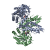
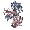
| Title | Crystal Structure of human O-GlcNAcase in complex with glycopeptide p53 | ||||||
 Components Components |
| ||||||
 Keywords Keywords |  HYDROLASE / human O-GlcNAcase / glycopeptide HYDROLASE / human O-GlcNAcase / glycopeptide | ||||||
| Function / homology |  Function and homology information Function and homology informationglycoprotein metabolic process / hyalurononglucosaminidase activity / N-acetylglucosamine metabolic process / glycoprotein catabolic process / protein O-GlcNAcase / : / : / [protein]-3-O-(N-acetyl-D-glucosaminyl)-L-serine/L-threonine O-N-acetyl-alpha-D-glucosaminase activity / protein O-linked glycosylation / protein deglycosylation ...glycoprotein metabolic process / hyalurononglucosaminidase activity / N-acetylglucosamine metabolic process / glycoprotein catabolic process / protein O-GlcNAcase / : / : / [protein]-3-O-(N-acetyl-D-glucosaminyl)-L-serine/L-threonine O-N-acetyl-alpha-D-glucosaminase activity / protein O-linked glycosylation / protein deglycosylation / Loss of function of TP53 in cancer due to loss of tetramerization ability / Regulation of TP53 Expression / signal transduction by p53 class mediator / negative regulation of G1 to G0 transition / negative regulation of glucose catabolic process to lactate via pyruvate / Transcriptional activation of cell cycle inhibitor p21 / regulation of intrinsic apoptotic signaling pathway by p53 class mediator / Activation of NOXA and translocation to mitochondria / negative regulation of pentose-phosphate shunt / ATP-dependent DNA/DNA annealing activity / negative regulation of helicase activity / regulation of cell cycle G2/M phase transition / intrinsic apoptotic signaling pathway in response to hypoxia / regulation of fibroblast apoptotic process / oxidative stress-induced premature senescence / oligodendrocyte apoptotic process / negative regulation of miRNA processing / positive regulation of thymocyte apoptotic process / glucose catabolic process to lactate via pyruvate / regulation of tissue remodeling / positive regulation of mitochondrial membrane permeability / negative regulation of mitophagy / positive regulation of programmed necrotic cell death / mRNA transcription / bone marrow development / circadian behavior / T cell proliferation involved in immune response / regulation of mitochondrial membrane permeability involved in apoptotic process / histone deacetylase regulator activity / RUNX3 regulates CDKN1A transcription / germ cell nucleus / regulation of DNA damage response, signal transduction by p53 class mediator / TP53 regulates transcription of additional cell cycle genes whose exact role in the p53 pathway remain uncertain / TP53 Regulates Transcription of Death Receptors and Ligands / Activation of PUMA and translocation to mitochondria / DNA damage response, signal transduction by p53 class mediator resulting in transcription of p21 class mediator / negative regulation of glial cell proliferation / Formation of Senescence-Associated Heterochromatin Foci (SAHF) / negative regulation of neuroblast proliferation / Regulation of TP53 Activity through Association with Co-factors / positive regulation of execution phase of apoptosis / mitochondrial DNA repair / T cell lineage commitment / negative regulation of DNA replication / ER overload response / B cell lineage commitment / thymocyte apoptotic process / positive regulation of cardiac muscle cell apoptotic process / TP53 regulates transcription of several additional cell death genes whose specific roles in p53-dependent apoptosis remain uncertain / TP53 Regulates Transcription of Caspase Activators and Caspases / cardiac septum morphogenesis / entrainment of circadian clock by photoperiod / PI5P Regulates TP53 Acetylation / Association of TriC/CCT with target proteins during biosynthesis / Zygotic genome activation (ZGA) / necroptotic process / rRNA transcription / positive regulation of release of cytochrome c from mitochondria / TP53 Regulates Transcription of Genes Involved in Cytochrome C Release / TFIID-class transcription factor complex binding / mitophagy / negative regulation of telomere maintenance via telomerase / SUMOylation of transcription factors / intrinsic apoptotic signaling pathway by p53 class mediator / general transcription initiation factor binding / neuroblast proliferation / cellular response to actinomycin D / Transcriptional Regulation by VENTX / response to X-ray / DNA damage response, signal transduction by p53 class mediator / replicative senescence / intrinsic apoptotic signaling pathway in response to endoplasmic reticulum stress / chromosome organization / gastrulation / cellular response to UV-C / response to inorganic substance / hematopoietic stem cell differentiation / negative regulation of reactive oxygen species metabolic process / intrinsic apoptotic signaling pathway in response to DNA damage by p53 class mediator / MDM2/MDM4 family protein binding / glial cell proliferation / positive regulation of RNA polymerase II transcription preinitiation complex assembly / embryonic organ development / Pyroptosis / cis-regulatory region sequence-specific DNA binding / hematopoietic progenitor cell differentiation / cellular response to glucose starvation / TP53 Regulates Transcription of Genes Involved in G1 Cell Cycle Arrest / somitogenesis / type II interferon-mediated signaling pathwaySimilarity search - Function | ||||||
| Biological species |  Homo sapiens (human) Homo sapiens (human) | ||||||
| Method | X-RAY DIFFRACTION / SYNCHROTRON / MOLECULAR REPLACEMENT / Resolution: 2.13 Å | ||||||
 Authors Authors | Li, B. / Jiang, J. | ||||||
| Funding support |  United States, 1items United States, 1items
| ||||||
 Citation Citation | Journal: Nat. Struct. Mol. Biol. / Year: 2017 Title: Structures of human O-GlcNAcase and its complexes reveal a new substrate recognition mode. Authors: Li, B. / Li, H. / Lu, L. / Jiang, J. | ||||||
| History |
|
- Structure visualization
| Structure viewer | Molecule: MolmilJmol/JSmol |
|---|
- Downloads & links
-Download
| PDBx/mmCIF format | 5un8.cif.gz | 389.2 KB | Display | PDBx/mmCIF format |
|---|---|---|---|---|
| PDB format | pdb5un8.ent.gz | 315 KB | Display | PDB format |
| PDBx/mmJSON format | 5un8.json.gz | Tree view | PDBx/mmJSON format | |
| Others |  Other downloads Other downloads |
-Validation report
| Arichive directory | https://data.pdbj.org/pub/pdb/validation_reports/un/5un8ftp://data.pdbj.org/pub/pdb/validation_reports/un/5un8 | HTTPS FTP |
|---|
-Related structure data
| Related structure data |  5tkeSC  5un9C C: citing same article ( S: Starting model for refinement |
|---|---|
| Similar structure data |


-Links
 PDBj
PDBj


- Assembly
| Deposited unit | 
| ||||||||
|---|---|---|---|---|---|---|---|---|---|
| 1 | 
| ||||||||
| 2 | 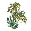
| ||||||||
| Unit cell |
|
-Components
| #1: Protein | / OGA / Beta-N-acetylglucosaminidase / Beta-N-acetylhexosaminidase / Beta-hexosaminidase / Meningioma- ...OGA / Beta-N-acetylglucosaminidase / Beta-N-acetylhexosaminidase / Beta-hexosaminidase / Meningioma-expressed antigen 5 / N-acetyl-beta-D-glucosaminidase / N-acetyl-beta-glucosaminidase / Nuclear cytoplasmic O-GlcNAcase and acetyltransferase / NCOAT Mass: 57822.582 Da / Num. of mol.: 4 / Fragment: UNP residues 60-400 and 553-704 Source method: isolated from a genetically manipulated source Source: (gene. exp.) Homo sapiens (human) / Gene: MGEA5, HEXC, KIAA0679, MEA5Production host: References: UniProt: O60502, protein O-GlcNAcase, Hydrolases; Glycosylases; Glycosidases, i.e. enzymes that hydrolyse O- and S-glycosyl compounds#2: Protein/peptide | Mass: 1196.308 Da / Num. of mol.: 4 / Source method: obtained synthetically / Details: Ser was O-GlcNAcylated; NAG stands for O-GlcNAc / Source: (synth.) Homo sapiens (human) / References: UniProt: P04637*PLUS#3: Sugar | ChemComp-NAG / N-Acetylglucosamine  Type: D-saccharide, beta linking / Mass: 221.208 Da / Num. of mol.: 4 Type: D-saccharide, beta linking / Mass: 221.208 Da / Num. of mol.: 4Source method: isolated from a genetically manipulated source Formula: C8H15NO6 #4: Water | ChemComp-HOH / | Water Mass: 18.015 Da / Num. of mol.: 1232 / Source method: isolated from a natural source / Formula: H2O Mass: 18.015 Da / Num. of mol.: 1232 / Source method: isolated from a natural source / Formula: H2O |
|---|
-Experimental details
-Experiment
| Experiment | Method: X-RAY DIFFRACTION / Number of used crystals: 1 |
|---|
- Sample preparation
| Crystal | Density Matthews: 2.93 Å3/Da / Density % sol: 58 % |
|---|---|
| Crystal grow | Temperature: 293 K / Method: evaporation Details: 0.032 M ammonium citrate tribasic (pH 7.0), 0.02 M MES monohydrate, 0.016 M imidazole, 0.002 M zinc sulfate heptahydrate, 0.128 M potassium thiocyanate, 12.8% w/v polyethylene glycol 3,350, ...Details: 0.032 M ammonium citrate tribasic (pH 7.0), 0.02 M MES monohydrate, 0.016 M imidazole, 0.002 M zinc sulfate heptahydrate, 0.128 M potassium thiocyanate, 12.8% w/v polyethylene glycol 3,350, 3.2% w/v polyethylene glycol monomethyl ether 2,000, and 5% w/v polyethylene glycol monomethyl ether 550. |
-Data collection
| Diffraction | Mean temperature: 80 K |
|---|---|
| Diffraction source | Source: SYNCHROTRON / Site: APS / Beamline: 21-ID-D / Wavelength: 0.978 Å |
| Detector | Type: MARMOSAIC 225 mm CCD / Detector: CCD / Date: Jun 4, 2016 |
| Radiation | Protocol: SINGLE WAVELENGTH / Monochromatic (M) / Laue (L): M / Scattering type: x-ray |
| Radiation wavelength | Wavelength: 0.978 Å / Relative weight: 1 |
| Reflection | Resolution: 2.13→148.24 Å / Num. obs: 139265 / % possible obs: 99.9 % / Redundancy: 4.9 % / Rpim(I) all: 0.042 / Net I/σ(I): 18.4 |
| Reflection shell | Resolution: 2.14→2.18 Å / Redundancy: 4.5 % / Mean I/σ(I) obs: 2 / Num. unique obs: 6934 / CC1/2: 0.781 / Rpim(I) all: 0.379 / % possible all: 100 |
- Processing
| Software |
| ||||||||||||||||||||||||||||||||||||||||||||||||||||||||||||||||||||||||||||||||||||||||||||||||||||||||||||||||||||||||||||||||||||||||||||||||||||||||||||||||||||||||||||||||||||||
|---|---|---|---|---|---|---|---|---|---|---|---|---|---|---|---|---|---|---|---|---|---|---|---|---|---|---|---|---|---|---|---|---|---|---|---|---|---|---|---|---|---|---|---|---|---|---|---|---|---|---|---|---|---|---|---|---|---|---|---|---|---|---|---|---|---|---|---|---|---|---|---|---|---|---|---|---|---|---|---|---|---|---|---|---|---|---|---|---|---|---|---|---|---|---|---|---|---|---|---|---|---|---|---|---|---|---|---|---|---|---|---|---|---|---|---|---|---|---|---|---|---|---|---|---|---|---|---|---|---|---|---|---|---|---|---|---|---|---|---|---|---|---|---|---|---|---|---|---|---|---|---|---|---|---|---|---|---|---|---|---|---|---|---|---|---|---|---|---|---|---|---|---|---|---|---|---|---|---|---|---|---|---|---|
| Refinement | Method to determine structure: MOLECULAR REPLACEMENT Starting model: 5TKE Resolution: 2.13→148.24 Å / Cor.coef. Fo:Fc: 0.961 / Cor.coef. Fo:Fc free: 0.94 / SU B: 4.878 / SU ML: 0.125 / Cross valid method: THROUGHOUT / ESU R: 0.195 / ESU R Free: 0.175 / Stereochemistry target values: MAXIMUM LIKELIHOOD
| ||||||||||||||||||||||||||||||||||||||||||||||||||||||||||||||||||||||||||||||||||||||||||||||||||||||||||||||||||||||||||||||||||||||||||||||||||||||||||||||||||||||||||||||||||||||
| Solvent computation | Ion probe radii: 0.8 Å / Shrinkage radii: 0.8 Å / VDW probe radii: 1.2 Å / Solvent model: MASK | ||||||||||||||||||||||||||||||||||||||||||||||||||||||||||||||||||||||||||||||||||||||||||||||||||||||||||||||||||||||||||||||||||||||||||||||||||||||||||||||||||||||||||||||||||||||
| Displacement parameters | Biso mean: 30.017 Å2
| ||||||||||||||||||||||||||||||||||||||||||||||||||||||||||||||||||||||||||||||||||||||||||||||||||||||||||||||||||||||||||||||||||||||||||||||||||||||||||||||||||||||||||||||||||||||
| Refinement step | Cycle: 1 / Resolution: 2.13→148.24 Å
| ||||||||||||||||||||||||||||||||||||||||||||||||||||||||||||||||||||||||||||||||||||||||||||||||||||||||||||||||||||||||||||||||||||||||||||||||||||||||||||||||||||||||||||||||||||||
| Refine LS restraints |
|