 Movie
Movie Controller

[English] 日本語
 Yorodumi
- PDB-5tvu: Crystal structure of mitochondrial Hsp90 (TRAP1) with ATP in abse... -
+ Open data

- Basic information
| Entry | Database: PDB / ID: 5tvu | ||||||
|---|---|---|---|---|---|---|---|
| Title | Crystal structure of mitochondrial Hsp90 (TRAP1) with ATP in absence of Mg | ||||||
 Components Components | TNF receptor-associated protein 1 | ||||||
 Keywords Keywords |  CHAPERONE / Hsp90 / ATP / TRAP1 / Structural Genomics / PSI-2 / Protein Structure Initiative CHAPERONE / Hsp90 / ATP / TRAP1 / Structural Genomics / PSI-2 / Protein Structure Initiative | ||||||
| Function / homology |  Function and homology information: / nucleic acid metabolic process / ATP-dependent protein folding chaperone / unfolded protein binding / protein folding / mitochondrial inner membrane / calcium ion binding / protein kinase binding / magnesium ion binding / ATP hydrolysis activity ...: / nucleic acid metabolic process / ATP-dependent protein folding chaperone / unfolded protein binding / protein folding / mitochondrial inner membrane / calcium ion binding / protein kinase binding / magnesium ion binding / ATP hydrolysis activity / ATP binding / identical protein binding Function and homology information: / nucleic acid metabolic process / ATP-dependent protein folding chaperone / unfolded protein binding / protein folding / mitochondrial inner membrane / calcium ion binding / protein kinase binding / magnesium ion binding / ATP hydrolysis activity ...: / nucleic acid metabolic process / ATP-dependent protein folding chaperone / unfolded protein binding / protein folding / mitochondrial inner membrane / calcium ion binding / protein kinase binding / magnesium ion binding / ATP hydrolysis activity / ATP binding / identical protein bindingSimilarity search - Function | ||||||
| Biological species |  Danio rerio (zebrafish) Danio rerio (zebrafish) | ||||||
| Method | X-RAY DIFFRACTION / SYNCHROTRON / MOLECULAR REPLACEMENT / Resolution: 3.5 Å | ||||||
 Authors Authors | Elnatan, D. / Betegon, M. / Agard, D.A. | ||||||
| Funding support |  United States, 1items United States, 1items
| ||||||
 Citation Citation | Journal: Elife / Year: 2017 Title: Symmetry broken and rebroken during the ATP hydrolysis cycle of the mitochondrial Hsp90 TRAP1. Authors: Elnatan, D. / Betegon, M. / Liu, Y. / Ramelot, T. / Kennedy, M.A. / Agard, D.A. | ||||||
| History |
|
- Structure visualization
| Structure viewer | Molecule: MolmilJmol/JSmol |
|---|
- Downloads & links
-Download
| PDBx/mmCIF format | 5tvu.cif.gz | 496.7 KB | Display | PDBx/mmCIF format |
|---|---|---|---|---|
| PDB format | pdb5tvu.ent.gz | 406.3 KB | Display | PDB format |
| PDBx/mmJSON format | 5tvu.json.gz | Tree view | PDBx/mmJSON format | |
| Others |  Other downloads Other downloads |
-Validation report
| Arichive directory | https://data.pdbj.org/pub/pdb/validation_reports/tv/5tvuftp://data.pdbj.org/pub/pdb/validation_reports/tv/5tvu | HTTPS FTP |
|---|
-Related structure data
| Related structure data |  5tthC  5tvwC 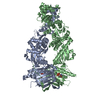 5tvxC  4ipeS C: citing same article ( S: Starting model for refinement |
|---|---|
| Similar structure data |


-Links
 PDBj
PDBj

- Assembly
| Deposited unit | 
| ||||||||||
|---|---|---|---|---|---|---|---|---|---|---|---|
| 1 |
| ||||||||||
| Unit cell |
|
-Components
| #1: Protein | Mass: 74635.625 Da / Num. of mol.: 2 / Fragment: UNP residues 73-719 Source method: isolated from a genetically manipulated source Source: (gene. exp.) Danio rerio (zebrafish) / Gene: trap1 / Variant: delta1-72 / Plasmid: pET151DTOPO / Production host:  Escherichia coli BL21(DE3) (bacteria) / Strain (production host): BL21(DE3)-RIL / References: UniProt: A8WFV1, UniProt: F1Q9X9*PLUS Escherichia coli BL21(DE3) (bacteria) / Strain (production host): BL21(DE3)-RIL / References: UniProt: A8WFV1, UniProt: F1Q9X9*PLUS#2: Chemical | Adenosine triphosphate  Mass: 507.181 Da / Num. of mol.: 2 / Source method: obtained synthetically / Formula: C10H16N5O13P3 / Comment: ATP, energy-carrying molecule*YM Mass: 507.181 Da / Num. of mol.: 2 / Source method: obtained synthetically / Formula: C10H16N5O13P3 / Comment: ATP, energy-carrying molecule*YM |
|---|
-Experimental details
-Experiment
| Experiment | Method: X-RAY DIFFRACTION / Number of used crystals: 1 |
|---|
- Sample preparation
| Crystal | Density Matthews: 2.31 Å3/Da / Density % sol: 46.72 % |
|---|---|
| Crystal grow | Temperature: 293.15 K / Method: vapor diffusion, hanging drop Details: 0.2 M NaK Tartrate, 20% (v/v) PEG3350, 36 mM hexamine cobalt, 5 mM ATP,5 mM EDTA |
-Data collection
| Diffraction | Mean temperature: 91.4 K |
|---|---|
| Diffraction source | Source: SYNCHROTRON / Site: ALS / Beamline: 8.3.1 / Wavelength: 1.1159 Å |
| Detector | Type: ADSC QUANTUM 315r / Detector: CCD / Date: Aug 1, 2015 |
| Radiation | Protocol: SINGLE WAVELENGTH / Monochromatic (M) / Laue (L): M / Scattering type: x-ray |
| Radiation wavelength | Wavelength: 1.1159 Å / Relative weight: 1 |
| Reflection | Resolution: 2.3→44.52 Å / Num. obs: 61835 / % possible obs: 93.4 % / Redundancy: 1.8 % / CC1/2: 0.734 / Net I/σ(I): 2.2 |
| Reflection shell | Resolution: 2.3→2.39 Å |
- Processing
| Software |
| ||||||||||||||||||||||||||||||||||||||||||||||||||||||||||||||||||||||||||||||||||||||||||||||||||||||||||||||||||||||||||||||||||||||||||||||||||||||||||||||||||||||||||||||||||||||||||||||||||||||||
|---|---|---|---|---|---|---|---|---|---|---|---|---|---|---|---|---|---|---|---|---|---|---|---|---|---|---|---|---|---|---|---|---|---|---|---|---|---|---|---|---|---|---|---|---|---|---|---|---|---|---|---|---|---|---|---|---|---|---|---|---|---|---|---|---|---|---|---|---|---|---|---|---|---|---|---|---|---|---|---|---|---|---|---|---|---|---|---|---|---|---|---|---|---|---|---|---|---|---|---|---|---|---|---|---|---|---|---|---|---|---|---|---|---|---|---|---|---|---|---|---|---|---|---|---|---|---|---|---|---|---|---|---|---|---|---|---|---|---|---|---|---|---|---|---|---|---|---|---|---|---|---|---|---|---|---|---|---|---|---|---|---|---|---|---|---|---|---|---|---|---|---|---|---|---|---|---|---|---|---|---|---|---|---|---|---|---|---|---|---|---|---|---|---|---|---|---|---|---|---|---|---|
| Refinement | Method to determine structure: MOLECULAR REPLACEMENT Starting model: 4IPE Resolution: 3.5→39.759 Å / SU ML: 0.44 / Cross valid method: FREE R-VALUE / σ(F): 1.34 / Phase error: 27.47 / Stereochemistry target values: ML
| ||||||||||||||||||||||||||||||||||||||||||||||||||||||||||||||||||||||||||||||||||||||||||||||||||||||||||||||||||||||||||||||||||||||||||||||||||||||||||||||||||||||||||||||||||||||||||||||||||||||||
| Solvent computation | Shrinkage radii: 0.9 Å / VDW probe radii: 1.11 Å / Solvent model: FLAT BULK SOLVENT MODEL | ||||||||||||||||||||||||||||||||||||||||||||||||||||||||||||||||||||||||||||||||||||||||||||||||||||||||||||||||||||||||||||||||||||||||||||||||||||||||||||||||||||||||||||||||||||||||||||||||||||||||
| Refinement step | Cycle: LAST / Resolution: 3.5→39.759 Å
| ||||||||||||||||||||||||||||||||||||||||||||||||||||||||||||||||||||||||||||||||||||||||||||||||||||||||||||||||||||||||||||||||||||||||||||||||||||||||||||||||||||||||||||||||||||||||||||||||||||||||
| Refine LS restraints |
| ||||||||||||||||||||||||||||||||||||||||||||||||||||||||||||||||||||||||||||||||||||||||||||||||||||||||||||||||||||||||||||||||||||||||||||||||||||||||||||||||||||||||||||||||||||||||||||||||||||||||
| LS refinement shell |
| ||||||||||||||||||||||||||||||||||||||||||||||||||||||||||||||||||||||||||||||||||||||||||||||||||||||||||||||||||||||||||||||||||||||||||||||||||||||||||||||||||||||||||||||||||||||||||||||||||||||||
| Refinement TLS params. | Method: refined / Refine-ID: X-RAY DIFFRACTION
| ||||||||||||||||||||||||||||||||||||||||||||||||||||||||||||||||||||||||||||||||||||||||||||||||||||||||||||||||||||||||||||||||||||||||||||||||||||||||||||||||||||||||||||||||||||||||||||||||||||||||
| Refinement TLS group |
|
