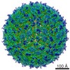
Entry Database : PDB / ID : 5iy3Title Zika Virus Non-structural Protein NS1 Genome polyprotein Keywords / Function / homology Function Domain/homology Component
/ / / / / / / / / / / / / / / / / / / / / / / / / / / / / / / / / / / / / / / / / / / / / / / / / / / / / / / / / / / / / / / / / / / / / / / / / / / / / / / / / / / / / / / / / / / / / / / / / / / / / / / / / / / / / / / / / / / / / / / / / / Biological species Method / / / Resolution : 2.2 Å Authors Song, H. / Qi, J. / Shi, Y. / Gao, G.F. Journal : Nat.Struct.Mol.Biol. / Year : 2016Title : Zika virus NS1 structure reveals diversity of electrostatic surfaces among flavivirusesAuthors : Song, H. / Qi, J. / Haywood, J. / Shi, Y. / Gao, G.F. History Deposition Mar 23, 2016 Deposition site / Processing site Revision 1.0 Apr 13, 2016 Provider / Type Revision 1.1 Apr 20, 2016 Group Revision 1.2 Apr 27, 2016 Group Revision 1.3 May 25, 2016 Group Revision 1.4 Nov 8, 2023 Group Data collection / Database references ... Data collection / Database references / Derived calculations / Refinement description Category chem_comp_atom / chem_comp_bond ... chem_comp_atom / chem_comp_bond / database_2 / pdbx_initial_refinement_model / pdbx_struct_oper_list Item / _database_2.pdbx_database_accession / _pdbx_struct_oper_list.symmetry_operation
Show all Show less
 Movie
Movie Controller


 Open data
 Basic information
 Components
 Keywords
 VIRAL PROTEIN /
  Function and homology information
 Authors
 Citation
 Structure visualization
 Downloads & links
 Other downloads
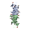


 PDBj
PDBj


 Assembly

 Mass: 18.015 Da / Num. of mol.: 79 / Source method: isolated from a natural source / Formula: H2O
 Sample preparation
 / Beamline: BL17U / Wavelength: 0.97915 Å
 Processing