 Movie
Movie Controller


+ Open data

- Basic information
| Entry | Database: PDB / ID: 5i9i | ||||||
|---|---|---|---|---|---|---|---|
| Title | Crystal structure of LP_PLA2 in complex with Darapladib | ||||||
 Components Components | Platelet-activating factor acetylhydrolase 1-alkyl-2-acetylglycerophosphocholine esterase 1-alkyl-2-acetylglycerophosphocholine esterase | ||||||
 Keywords Keywords | HYDROLASE/HYDROLASE INHIBITOR / Lp_pla2 inhibitor / HYDROLASE-HYDROLASE INHIBITOR complex | ||||||
| Function / homology |  Function and homology information Function and homology informationplasma lipoprotein particle oxidation / platelet activating factor catabolic process / calcium-independent phospholipase A2 activity / 1-alkyl-2-acetylglycerophosphocholine esterase / platelet activating factor metabolic process / 1-alkyl-2-acetylglycerophosphocholine esterase activity / lipid oxidation / phosphatidylcholine catabolic process / low-density lipoprotein particle / high-density lipoprotein particle ...plasma lipoprotein particle oxidation / platelet activating factor catabolic process / calcium-independent phospholipase A2 activity / 1-alkyl-2-acetylglycerophosphocholine esterase / platelet activating factor metabolic process / 1-alkyl-2-acetylglycerophosphocholine esterase activity / lipid oxidation / phosphatidylcholine catabolic process / low-density lipoprotein particle / high-density lipoprotein particle / low-density lipoprotein particle remodeling / positive regulation of monocyte chemotaxis / peptide hormone processing / hydrolase activity, acting on ester bonds / Synthesis, secretion, and deacylation of Ghrelin / phospholipid binding / positive regulation of inflammatory response / extracellular regionSimilarity search - Function | ||||||
| Biological species |  Homo sapiens (human) Homo sapiens (human) | ||||||
| Method | X-RAY DIFFRACTION / SYNCHROTRON / MOLECULAR REPLACEMENT / molecular replacement / Resolution: 2.7 Å | ||||||
 Authors Authors | Liu, Q.F. / Xu, Y.C. | ||||||
 Citation Citation | Journal: J.Med.Chem. / Year: 2016 Title: Structural and Thermodynamic Characterization of Protein-Ligand Interactions Formed between Lipoprotein-Associated Phospholipase A2 and Inhibitors Authors: Liu, Q.F. / Chen, X.D. / Chen, W.Y. / Yuan, X.J. / Su, H.X. / Shen, J.H. / Xu, Y.C. | ||||||
| History |
|
- Structure visualization
| Structure viewer | Molecule: MolmilJmol/JSmol |
|---|
- Downloads & links
-Download
| PDBx/mmCIF format | 5i9i.cif.gz | 155.4 KB | Display | PDBx/mmCIF format |
|---|---|---|---|---|
| PDB format | pdb5i9i.ent.gz | 119.1 KB | Display | PDB format |
| PDBx/mmJSON format | 5i9i.json.gz | Tree view | PDBx/mmJSON format | |
| Others |  Other downloads Other downloads |
-Validation report
| Arichive directory | https://data.pdbj.org/pub/pdb/validation_reports/i9/5i9iftp://data.pdbj.org/pub/pdb/validation_reports/i9/5i9i | HTTPS FTP |
|---|
-Related structure data
| Related structure data |  5i8pC  3d59S C: citing same article ( S: Starting model for refinement |
|---|---|
| Similar structure data |


-Links
 PDBj
PDBj


- Assembly
| Deposited unit | 
| ||||||||
|---|---|---|---|---|---|---|---|---|---|
| 1 | 
| ||||||||
| 2 | 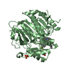
| ||||||||
| Unit cell |
|
-Components
| #1: Protein | 1-alkyl-2-acetylglycerophosphocholine esterase / PAF acetylhydrolase / 1-alkyl-2-acetylglycerophosphocholine esterase / 2-acetyl-1- ...PAF acetylhydrolase / 1-alkyl-2-acetylglycerophosphocholine esterase / 2-acetyl-1-alkylglycerophosphocholine esterase / Group-VIIA phospholipase A2 / gVIIA-PLA2 / LDL-associated phospholipase A2 / LDL-PLA(2) / PAF 2-acylhydrolase Mass: 43905.816 Da / Num. of mol.: 2 / Fragment: UNP residues 47-429 Source method: isolated from a genetically manipulated source Source: (gene. exp.) Homo sapiens (human) / Gene: PLA2G7, PAFAH / Plasmid: pGEX-6P-1 / Production host:  Escherichia coli (E. coli) / Strain (production host): Rosetta2 pLysS Escherichia coli (E. coli) / Strain (production host): Rosetta2 pLysSReferences: UniProt: Q13093, 1-alkyl-2-acetylglycerophosphocholine esterase#2: Chemical | Darapladib  Mass: 666.771 Da / Num. of mol.: 2 / Source method: obtained synthetically / Formula: C36H38F4N4O2S / Comment: inhibitor*YM Mass: 666.771 Da / Num. of mol.: 2 / Source method: obtained synthetically / Formula: C36H38F4N4O2S / Comment: inhibitor*YM#3: Chemical | Sulfate  Mass: 96.063 Da / Num. of mol.: 2 / Source method: isolated from a natural source / Formula: SO4 Mass: 96.063 Da / Num. of mol.: 2 / Source method: isolated from a natural source / Formula: SO4#4: Water | ChemComp-HOH / | Water Mass: 18.015 Da / Num. of mol.: 6 / Source method: isolated from a natural source / Formula: H2O Mass: 18.015 Da / Num. of mol.: 6 / Source method: isolated from a natural source / Formula: H2O |
|---|
-Experimental details
-Experiment
| Experiment | Method: X-RAY DIFFRACTION / Number of used crystals: 1 |
|---|
- Sample preparation
| Crystal | Density Matthews: 2.37 Å3/Da / Density % sol: 48.06 % |
|---|---|
| Crystal grow | Temperature: 293 K / Method: vapor diffusion, hanging drop / pH: 6.6 Details: 0.1M MOPS pH 6.6, 0.4M Li2SO4, 27% (w/v) (NH4)2SO4, 1M Na-Ac, 1.4% 1,4-butanediol |
-Data collection
| Diffraction | Mean temperature: 100 K | ||||||||||||||||||||||||||||||||||||||||||||||||||||||||||||||||||||||||||||||||||||||||||||||||||||||||||||||||||||||||||||||
|---|---|---|---|---|---|---|---|---|---|---|---|---|---|---|---|---|---|---|---|---|---|---|---|---|---|---|---|---|---|---|---|---|---|---|---|---|---|---|---|---|---|---|---|---|---|---|---|---|---|---|---|---|---|---|---|---|---|---|---|---|---|---|---|---|---|---|---|---|---|---|---|---|---|---|---|---|---|---|---|---|---|---|---|---|---|---|---|---|---|---|---|---|---|---|---|---|---|---|---|---|---|---|---|---|---|---|---|---|---|---|---|---|---|---|---|---|---|---|---|---|---|---|---|---|---|---|---|
| Diffraction source | Source: SYNCHROTRON / Site: SSRF  / Beamline: BL17U / Wavelength: 0.9791 Å / Beamline: BL17U / Wavelength: 0.9791 Å | ||||||||||||||||||||||||||||||||||||||||||||||||||||||||||||||||||||||||||||||||||||||||||||||||||||||||||||||||||||||||||||||
| Detector | Type: ADSC QUANTUM 315r / Detector: CCD / Date: Dec 15, 2015 | ||||||||||||||||||||||||||||||||||||||||||||||||||||||||||||||||||||||||||||||||||||||||||||||||||||||||||||||||||||||||||||||
| Radiation | Protocol: SINGLE WAVELENGTH / Monochromatic (M) / Laue (L): M / Scattering type: x-ray | ||||||||||||||||||||||||||||||||||||||||||||||||||||||||||||||||||||||||||||||||||||||||||||||||||||||||||||||||||||||||||||||
| Radiation wavelength | Wavelength: 0.9791 Å / Relative weight: 1 | ||||||||||||||||||||||||||||||||||||||||||||||||||||||||||||||||||||||||||||||||||||||||||||||||||||||||||||||||||||||||||||||
| Reflection | Resolution: 2.7→44.064 Å / Num. obs: 22260 / % possible obs: 95.5 % / Observed criterion σ(I): -3 / Redundancy: 1.96 % / Biso Wilson estimate: 34.72 Å2 / CC1/2: 0.985 / Rmerge(I) obs: 0.094 / Net I/σ(I): 8.59 | ||||||||||||||||||||||||||||||||||||||||||||||||||||||||||||||||||||||||||||||||||||||||||||||||||||||||||||||||||||||||||||||
| Reflection shell |
|
-Phasing
| Phasing | Method: molecular replacement | |||||||||
|---|---|---|---|---|---|---|---|---|---|---|
| Phasing MR |
|
- Processing
| Software |
| ||||||||||||||||||||||||||||||||||||||||||||||||||||||||||||||||||||||
|---|---|---|---|---|---|---|---|---|---|---|---|---|---|---|---|---|---|---|---|---|---|---|---|---|---|---|---|---|---|---|---|---|---|---|---|---|---|---|---|---|---|---|---|---|---|---|---|---|---|---|---|---|---|---|---|---|---|---|---|---|---|---|---|---|---|---|---|---|---|---|---|
| Refinement | Method to determine structure: MOLECULAR REPLACEMENT Starting model: 3D59 Resolution: 2.7→44.064 Å / SU ML: 0.36 / Cross valid method: FREE R-VALUE / σ(F): 0.93 / Phase error: 26.34
| ||||||||||||||||||||||||||||||||||||||||||||||||||||||||||||||||||||||
| Solvent computation | Shrinkage radii: 0.9 Å / VDW probe radii: 1.11 Å | ||||||||||||||||||||||||||||||||||||||||||||||||||||||||||||||||||||||
| Displacement parameters | Biso max: 74.01 Å2 / Biso mean: 29.1156 Å2 / Biso min: 8.51 Å2 | ||||||||||||||||||||||||||||||||||||||||||||||||||||||||||||||||||||||
| Refinement step | Cycle: final / Resolution: 2.7→44.064 Å
| ||||||||||||||||||||||||||||||||||||||||||||||||||||||||||||||||||||||
| Refine LS restraints |
| ||||||||||||||||||||||||||||||||||||||||||||||||||||||||||||||||||||||
| LS refinement shell | Refine-ID: X-RAY DIFFRACTION / Total num. of bins used: 9
|
