 Movie
Movie Controller

[English] 日本語
 Yorodumi
- PDB-5grk: Crystal structure of Uracil DNA glycosylase -Xanthine complex fro... -
+ Open data

- Basic information
| Entry | Database: PDB / ID: 5grk | ||||||
|---|---|---|---|---|---|---|---|
| Title | Crystal structure of Uracil DNA glycosylase -Xanthine complex from Bradyrhizobium diazoefficiens | ||||||
 Components Components | Blr0248 protein | ||||||
 Keywords Keywords |  HYDROLASE / Uracil DNA glycosylase (UDG) Bradyrhizobium diazoefficiens / nitrogen fixing symbiont / DNA repair / Xanthine. HYDROLASE / Uracil DNA glycosylase (UDG) Bradyrhizobium diazoefficiens / nitrogen fixing symbiont / DNA repair / Xanthine. | ||||||
| Function / homology | Uracil-DNA glycosylase-like domain superfamily / XANTHINE / Blr0248 protein Function and homology information Function and homology information | ||||||
| Biological species |  Bradyrhizobium diazoefficiens USDA 110 (bacteria) Bradyrhizobium diazoefficiens USDA 110 (bacteria) | ||||||
| Method | X-RAY DIFFRACTION / SYNCHROTRON / Resolution: 2.804 Å | ||||||
 Authors Authors | Patil, V.V. / Ullas, V.C. / Ahn, W. / Varshney, U. / Woo, E. | ||||||
 Citation Citation | Journal: Nucleic Acids Res. / Year: 2017 Title: Uracil DNA glycosylase (UDG) activities in Bradyrhizobium diazoefficiens: characterization of a new class of UDG with broad substrate specificity Authors: Chembazhi, U.V. / Patil, V.V. / Sah, S. / Reeve, W. / Tiwari, R.P. / Woo, E. / Varshney, U. #1: Journal: NUCLEIC ACIDS RES. / Year: 2015 Title: A unique uracil-DNA binding protein of the uracil DNA glycosylase superfamily Authors: Sang, P.B. / Srinath, T. / Patil, A.G. / Woo, E. / Varshney, U. | ||||||
| History |
|
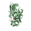
- Structure visualization
| Structure viewer | Molecule: MolmilJmol/JSmol |
|---|
- Downloads & links
-Download
| PDBx/mmCIF format | 5grk.cif.gz | 382.9 KB | Display | PDBx/mmCIF format |
|---|---|---|---|---|
| PDB format | pdb5grk.ent.gz | 330.3 KB | Display | PDB format |
| PDBx/mmJSON format | 5grk.json.gz | Tree view | PDBx/mmJSON format | |
| Others |  Other downloads Other downloads |
-Validation report
| Arichive directory | https://data.pdbj.org/pub/pdb/validation_reports/gr/5grkftp://data.pdbj.org/pub/pdb/validation_reports/gr/5grk | HTTPS FTP |
|---|
-Related structure data


-Links
 PDBj
PDBj
- Assembly
| Deposited unit | 
| ||||||||
|---|---|---|---|---|---|---|---|---|---|
| 1 | 
| ||||||||
| 2 | 
| ||||||||
| Unit cell |
|
-Components
| #1: Protein | Mass: 29725.795 Da / Num. of mol.: 4 Source method: isolated from a genetically manipulated source Source: (gene. exp.) Bradyrhizobium diazoefficiens USDA 110 (bacteria)Strain: USDA 110 / Gene: blr0248 / Production host: Escherichia coli (E. coli) / References: UniProt: Q89XR0#2: Chemical | ChemComp-XAN / Xanthine  Mass: 152.111 Da / Num. of mol.: 4 / Source method: obtained synthetically / Formula: C5H4N4O2 Mass: 152.111 Da / Num. of mol.: 4 / Source method: obtained synthetically / Formula: C5H4N4O2 |
|---|
-Experimental details
-Experiment
| Experiment | Method: X-RAY DIFFRACTION / Number of used crystals: 1 |
|---|
- Sample preparation
| Crystal | Density Matthews: 3.38 Å3/Da / Density % sol: 63.6 % |
|---|---|
| Crystal grow | Temperature: 291 K / Method: vapor diffusion, sitting drop / pH: 4 Details: 20% PEG3350, 200 mM sodium citrate, 100 mM sodium citrate/citric acid |
-Data collection
| Diffraction | Mean temperature: 93 K |
|---|---|
| Diffraction source | Source: SYNCHROTRON / Site: PAL/PLS  / Beamline: 5C (4A) / Wavelength: 0.97934 Å / Beamline: 5C (4A) / Wavelength: 0.97934 Å |
| Detector | Type: ADSC QUANTUM 270 / Detector: CCD / Date: Feb 26, 2016 |
| Radiation | Protocol: SINGLE WAVELENGTH / Monochromatic (M) / Laue (L): M / Scattering type: x-ray |
| Radiation wavelength | Wavelength: 0.97934 Å / Relative weight: 1 |
| Reflection | Resolution: 2.804→47.174 Å / Num. obs: 40422 / % possible obs: 99.91 % / Redundancy: 13.2 % / Net I/σ(I): 15.13 |
| Reflection shell | Resolution: 2.8→2.85 Å |
- Processing
| Software | Name: PHENIX / Version: 1.9_1692 / Classification: refinement | |||||||||||||||||||||||||||||||||||||||||||||||||||||||||||||||||||||||||||||||||||||||||||||||||||||||||
|---|---|---|---|---|---|---|---|---|---|---|---|---|---|---|---|---|---|---|---|---|---|---|---|---|---|---|---|---|---|---|---|---|---|---|---|---|---|---|---|---|---|---|---|---|---|---|---|---|---|---|---|---|---|---|---|---|---|---|---|---|---|---|---|---|---|---|---|---|---|---|---|---|---|---|---|---|---|---|---|---|---|---|---|---|---|---|---|---|---|---|---|---|---|---|---|---|---|---|---|---|---|---|---|---|---|---|
| Refinement | Resolution: 2.804→47.174 Å / SU ML: 0.33 / Cross valid method: FREE R-VALUE / σ(F): 1.37 / Phase error: 25.75 / Stereochemistry target values: ML
| |||||||||||||||||||||||||||||||||||||||||||||||||||||||||||||||||||||||||||||||||||||||||||||||||||||||||
| Solvent computation | Shrinkage radii: 0.9 Å / VDW probe radii: 1.11 Å / Solvent model: FLAT BULK SOLVENT MODEL | |||||||||||||||||||||||||||||||||||||||||||||||||||||||||||||||||||||||||||||||||||||||||||||||||||||||||
| Refinement step | Cycle: LAST / Resolution: 2.804→47.174 Å
| |||||||||||||||||||||||||||||||||||||||||||||||||||||||||||||||||||||||||||||||||||||||||||||||||||||||||
| Refine LS restraints |
| |||||||||||||||||||||||||||||||||||||||||||||||||||||||||||||||||||||||||||||||||||||||||||||||||||||||||
| LS refinement shell |
|
