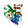
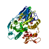
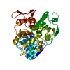
[English] 日本語
Yorodumi
- PDB-5gjc: Zika virus NS3 helicase in complex with ATP -

+
Open data


ID or keywords:

Loading...

-
Basic information

Entry
Database: PDB / ID: 5gjc
TitleZika virus NS3 helicase in complex with ATP
ComponentsNS3 helicase
KeywordsHYDROLASE / zika virus / helicase / ATP / Mn ion
Function / homology
Function and homology information


symbiont-mediated suppression of host JAK-STAT cascade via inhibition of STAT2 activity / ribonucleoside triphosphate phosphatase activity / double-stranded RNA binding / viral capsid / mRNA (nucleoside-2'-O-)-methyltransferase activity / mRNA 5'-cap (guanine-N7-)-methyltransferase activity / RNA helicase activity / host cell endoplasmic reticulum membrane / protein dimerization activity / symbiont-mediated suppression of host type I interferon-mediated signaling pathway ...symbiont-mediated suppression of host JAK-STAT cascade via inhibition of STAT2 activity / ribonucleoside triphosphate phosphatase activity / double-stranded RNA binding / viral capsid / mRNA (nucleoside-2'-O-)-methyltransferase activity / mRNA 5'-cap (guanine-N7-)-methyltransferase activity / RNA helicase activity / host cell endoplasmic reticulum membrane / protein dimerization activity / symbiont-mediated suppression of host type I interferon-mediated signaling pathway / symbiont entry into host cell / viral RNA genome replication / RNA-dependent RNA polymerase activity / serine-type endopeptidase activity / fusion of virus membrane with host endosome membrane / host cell nucleus / virion attachment to host cell / virion membrane / structural molecule activity / proteolysis / extracellular region / ATP binding / membrane / metal ion binding
Similarity search - Function
: / Flavivirus envelope glycoprotein E, stem/anchor domain / Flavivirus capsid protein C superfamily / RNA-directed RNA polymerase, thumb domain, Flavivirus / Flavivirus RNA-directed RNA polymerase, thumb domain / Flavivirus non-structural protein NS2B / : / Flavivirus NS3 helicase, C-terminal helical domain / Genome polyprotein, Flavivirus / Flavivirus non-structural protein NS4A ...: / Flavivirus envelope glycoprotein E, stem/anchor domain / Flavivirus capsid protein C superfamily / RNA-directed RNA polymerase, thumb domain, Flavivirus / Flavivirus RNA-directed RNA polymerase, thumb domain / Flavivirus non-structural protein NS2B / : / Flavivirus NS3 helicase, C-terminal helical domain / Genome polyprotein, Flavivirus / Flavivirus non-structural protein NS4A / Flavivirus non-structural protein NS2B / Flavivirus capsid protein C / Flavivirus non-structural protein NS4B / mRNA cap 0/1 methyltransferase / Flavivirus capsid protein C / Flavivirus non-structural protein NS4B / Flavivirus non-structural protein NS4A / Flavivirus NS2B domain profile. / mRNA cap 0 and cap 1 methyltransferase (EC 2.1.1.56 and EC 2.1.1.57) domain profile. / Flavivirus non-structural protein NS2A / Flavivirus non-structural protein NS2A / Flavivirus NS3, petidase S7 / Peptidase S7, Flavivirus NS3 serine protease / Flavivirus NS3 protease (NS3pro) domain profile. / Envelope glycoprotein M, flavivirus / Flavivirus envelope glycoprotein M / RNA-directed RNA polymerase, flavivirus / Flavivirus RNA-directed RNA polymerase, fingers and palm domains / Flavivirus non-structural Protein NS1 / Flavivirus non-structural protein NS1 / Envelope glycoprotein M superfamily, flavivirus / Flavivirus polyprotein propeptide / Flavivirus polyprotein propeptide superfamily / Flavivirus polyprotein propeptide / Flaviviral glycoprotein E, central domain, subdomain 1 / Flaviviral glycoprotein E, central domain, subdomain 2 / Flavivirus envelope glycoprotein E, Stem/Anchor domain / Flavivirus glycoprotein E, immunoglobulin-like domain / Flavivirus envelope glycoprotein E, Stem/Anchor domain superfamily / Flavivirus glycoprotein, immunoglobulin-like domain / Flavivirus glycoprotein central and dimerisation domain / Flavivirus glycoprotein, central and dimerisation domains / Ribosomal RNA methyltransferase, FtsJ domain / FtsJ-like methyltransferase / Flavivirus/Alphavirus glycoprotein, immunoglobulin-like domain superfamily / Flavivirus glycoprotein, central and dimerisation domain superfamily / Flaviviral glycoprotein E, dimerisation domain / DEAD box, Flavivirus / Flavivirus DEAD domain / helicase superfamily c-terminal domain / Immunoglobulin E-set / Superfamilies 1 and 2 helicase C-terminal domain profile. / Superfamilies 1 and 2 helicase ATP-binding type-1 domain profile. / DEAD-like helicases superfamily / Helicase, C-terminal / Helicase superfamily 1/2, ATP-binding domain / P-loop containing nucleotide triphosphate hydrolases / RNA-directed RNA polymerase, catalytic domain / RdRp of positive ssRNA viruses catalytic domain profile. / S-adenosyl-L-methionine-dependent methyltransferase superfamily / Peptidase S1, PA clan / DNA/RNA polymerase superfamily / P-loop containing nucleoside triphosphate hydrolase / Rossmann fold / 3-Layer(aba) Sandwich / Alpha Beta
Similarity search - Domain/homology
ADENOSINE-5'-TRIPHOSPHATE / : / Genome polyprotein / Genome polyprotein
Similarity search - Component
Biological speciesZika virus
MethodX-RAY DIFFRACTION / SYNCHROTRON / MOLECULAR REPLACEMENT / Resolution: 2.204 Å
AuthorsTian, H.L. / Ji, X.Y. / Yang, X.Y. / Zhang, Z.X. / Lu, Z.K. / Yang, K.L. / Chen, C. / Zhao, Q. / Chi, H. / Mu, Z.Y. ...Tian, H.L. / Ji, X.Y. / Yang, X.Y. / Zhang, Z.X. / Lu, Z.K. / Yang, K.L. / Chen, C. / Zhao, Q. / Chi, H. / Mu, Z.Y. / Xie, W. / Wang, Z.F. / Lou, H.Q. / Yang, H.T. / Rao, Z.H.
CitationJournal: Protein Cell / Year: 2016
Title: Structural basis of Zika virus helicase in recognizing its substrates
Authors: Tian, H.L. / Ji, X.Y. / Yang, X.Y. / Zhang, Z.X. / Lu, Z.K. / Yang, K.L. / Chen, C. / Zhao, Q. / Chi, H. / Mu, Z.Y. / Xie, W. / Wang, Z.F. / Lou, H.Q. / Yang, H.T. / Rao, Z.H.
History
DepositionJun 28, 2016Deposition site: PDBJ / Processing site: PDBJ
Revision 1.0Jul 20, 2016Provider: repository / Type: Initial release
Revision 1.1Aug 10, 2016Group: Database references
Revision 1.2Aug 31, 2016Group: Database references
Revision 1.3Nov 8, 2023Group: Data collection / Database references ...Data collection / Database references / Derived calculations / Refinement description
Category: chem_comp_atom / chem_comp_bond ...chem_comp_atom / chem_comp_bond / database_2 / pdbx_initial_refinement_model / pdbx_struct_conn_angle / pdbx_struct_oper_list / struct_conn
Item: _database_2.pdbx_DOI / _database_2.pdbx_database_accession ..._database_2.pdbx_DOI / _database_2.pdbx_database_accession / _pdbx_struct_conn_angle.ptnr1_auth_seq_id / _pdbx_struct_conn_angle.ptnr3_auth_seq_id / _pdbx_struct_conn_angle.value / _pdbx_struct_oper_list.symmetry_operation / _struct_conn.pdbx_dist_value / _struct_conn.ptnr2_auth_seq_id

-
Structure visualization

Structure viewerMolecule:
MolmilJmol/JSmol

Downloads & links

-
Assembly

Deposited unit
A: NS3 helicase
hetero molecules


Theoretical massNumber of molelcules
Total (without water)50,1983
Polymers49,6361
Non-polymers5622
Water1,928107
1


  • Idetical with deposited unit
  • defined by author&software
TypeNameSymmetry operationNumber
identity operation1_555x,y,z1
Buried area900 Å2
ΔGint-9 kcal/mol
Surface area19530 Å2
MethodPISA
Unit cell
Length a, b, c (Å)51.595, 74.167, 57.740
Angle α, β, γ (deg.)90.00, 92.21, 90.00
Int Tables number4
Space group name H-MP1211

-
Components

#1: Protein NS3 helicase


Mass: 49635.598 Da / Num. of mol.: 1 / Fragment: UNP residues 1682-2119
Source method: isolated from a genetically manipulated source
Source: (gene. exp.) Zika virus (strain Mr 766) / Strain: Mr 766 / Production host: Escherichia coli (E. coli) / References: UniProt: A0A142DS38, UniProt: A0A0U4DG08*PLUS
#2: Chemical ChemComp-MN / MANGANESE (II) ION


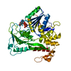
Mass: 54.938 Da / Num. of mol.: 1 / Source method: obtained synthetically / Formula: Mn
#3: Chemical ChemComp-ATP / ADENOSINE-5'-TRIPHOSPHATE / Adenosine triphosphate


Mass: 507.181 Da / Num. of mol.: 1 / Source method: obtained synthetically / Formula: C10H16N5O13P3 / Comment: ATP, energy-carrying molecule*YM
#4: Water ChemComp-HOH / water / Water


Mass: 18.015 Da / Num. of mol.: 107 / Source method: isolated from a natural source / Formula: H2O

-
Experimental details

-
Experiment

ExperimentMethod: X-RAY DIFFRACTION / Number of used crystals: 1

-
Sample preparation

CrystalDensity Matthews: 2.22 Å3/Da / Density % sol: 44.7 %
Crystal growTemperature: 291 K / Method: microbatch / pH: 7.4
Details: 0.1M HEPES(pH 7.4), 9% (w/v) polyethylene glycol 3350

-
Data collection

DiffractionMean temperature: 100 K
Diffraction sourceSource: SYNCHROTRON / Site: SSRF / Beamline: BL19U1 / Wavelength: 0.97853 Å
DetectorType: DECTRIS PILATUS3 S 6M / Detector: PIXEL / Date: Apr 12, 2016
RadiationProtocol: SINGLE WAVELENGTH / Monochromatic (M) / Laue (L): M / Scattering type: x-ray
Radiation wavelengthWavelength: 0.97853 Å / Relative weight: 1
ReflectionResolution: 2.2→40 Å / Num. obs: 42724 / % possible obs: 99.3 % / Redundancy: 6.7 % / Rmerge(I) obs: 0.083 / Net I/σ(I): 22.8
Reflection shellResolution: 2.2→2.24 Å / Rmerge(I) obs: 0.942

-
Processing

Software
NameVersionClassification
PHENIX1.9_1692refinement
HKL-3000data reduction
HKL-3000data scaling
PHASERphasing
RefinementMethod to determine structure: MOLECULAR REPLACEMENT
Starting model: 5JMT
Resolution: 2.204→37.729 Å / SU ML: 0.3 / Cross valid method: FREE R-VALUE / σ(F): 1.36 / Phase error: 29.12
Details: THE STRUCTURE FACTOR FILE CONTAINS FRIEDEL PAIRS IN F_PLUS/MINUS AND I_PLUS/MINUS COLUMNS.
RfactorNum. reflection% reflection
Rfree0.2474 2043 4.78 %
Rwork0.197 --
obs0.1995 42723 98.97 %
Solvent computationShrinkage radii: 0.9 Å / VDW probe radii: 1.11 Å
Refinement stepCycle: LAST / Resolution: 2.204→37.729 Å
ProteinNucleic acidLigandSolventTotal
Num. atoms3396 0 32 107 3535
Refine LS restraints
Refine-IDTypeDev idealNumber
X-RAY DIFFRACTIONf_bond_d0.0043502
X-RAY DIFFRACTIONf_angle_d0.9144750
X-RAY DIFFRACTIONf_dihedral_angle_d15.9161315
X-RAY DIFFRACTIONf_chiral_restr0.032524
X-RAY DIFFRACTIONf_plane_restr0.004609
LS refinement shell
Resolution (Å)Rfactor RfreeNum. reflection RfreeRfactor RworkNum. reflection RworkRefine-ID% reflection obs (%)
2.2044-2.25570.38421310.28392607X-RAY DIFFRACTION94
2.2557-2.31210.30471480.26772733X-RAY DIFFRACTION100
2.3121-2.37460.3142990.2452724X-RAY DIFFRACTION100
2.3746-2.44450.28851190.23922758X-RAY DIFFRACTION100
2.4445-2.52330.23071230.22132687X-RAY DIFFRACTION97
2.5233-2.61350.2771190.22372762X-RAY DIFFRACTION100
2.6135-2.71810.30121540.23282693X-RAY DIFFRACTION99
2.7181-2.84180.29121190.21942753X-RAY DIFFRACTION100
2.8418-2.99150.2891410.22082722X-RAY DIFFRACTION100
2.9915-3.17890.28051490.21822746X-RAY DIFFRACTION100
3.1789-3.42420.2371580.21262690X-RAY DIFFRACTION99
3.4242-3.76850.2251440.19212725X-RAY DIFFRACTION100
3.7685-4.31310.24881710.17672708X-RAY DIFFRACTION100
4.3131-5.43150.21461380.16842675X-RAY DIFFRACTION98
5.4315-37.73440.21371300.16992697X-RAY DIFFRACTION98

+
About Yorodumi

-
News

-
Feb 9, 2022. New format data for meta-information of EMDB entries

New format data for meta-information of EMDB entries

  • Version 3 of the EMDB header file is now the official format.
  • The previous official version 1.9 will be removed from the archive.

Related info.:EMDB header

External links:wwPDB to switch to version 3 of the EMDB data model

-
Aug 12, 2020. Covid-19 info

Covid-19 info

URL: https://pdbj.org/emnavi/covid19.php

New page: Covid-19 featured information page in EM Navigator.

Related info.:Covid-19 info / Mar 5, 2020. Novel coronavirus structure data

+
Mar 5, 2020. Novel coronavirus structure data

Novel coronavirus structure data

Related info.:Yorodumi Speices / Aug 12, 2020. Covid-19 info

External links:COVID-19 featured content - PDBj / Molecule of the Month (242):Coronavirus Proteases

+
Jan 31, 2019. EMDB accession codes are about to change! (news from PDBe EMDB page)

EMDB accession codes are about to change! (news from PDBe EMDB page)

  • The allocation of 4 digits for EMDB accession codes will soon come to an end. Whilst these codes will remain in use, new EMDB accession codes will include an additional digit and will expand incrementally as the available range of codes is exhausted. The current 4-digit format prefixed with “EMD-” (i.e. EMD-XXXX) will advance to a 5-digit format (i.e. EMD-XXXXX), and so on. It is currently estimated that the 4-digit codes will be depleted around Spring 2019, at which point the 5-digit format will come into force.
  • The EM Navigator/Yorodumi systems omit the EMD- prefix.

Related info.:Q: What is EMD? / ID/Accession-code notation in Yorodumi/EM Navigator

External links:EMDB Accession Codes are Changing Soon! / Contact to PDBj

+
Jul 12, 2017. Major update of PDB

Major update of PDB

  • wwPDB released updated PDB data conforming to the new PDBx/mmCIF dictionary.
  • This is a major update changing the version number from 4 to 5, and with Remediation, in which all the entries are updated.
  • In this update, many items about electron microscopy experimental information are reorganized (e.g. em_software).
  • Now, EM Navigator and Yorodumi are based on the updated data.

External links:wwPDB Remediation / Enriched Model Files Conforming to OneDep Data Standards Now Available in the PDB FTP Archive

-
Yorodumi

Thousand views of thousand structures

  • Yorodumi is a browser for structure data from EMDB, PDB, SASBDB, etc.
  • This page is also the successor to EM Navigator detail page, and also detail information page/front-end page for Omokage search.
  • The word "yorodu" (or yorozu) is an old Japanese word meaning "ten thousand". "mi" (miru) is to see.

Related info.:EMDB / PDB / SASBDB / Comparison of 3 databanks / Yorodumi Search / Aug 31, 2016. New EM Navigator & Yorodumi / Yorodumi Papers / Jmol/JSmol / Function and homology information / Changes in new EM Navigator and Yorodumi

Read more