Mass: 55757.812 Da / Num. of mol.: 1 / Fragment: CATALYTIC DOMAIN, RESIDUES 1-2,23-503 Source method: isolated from a genetically manipulated source Source: (gene. exp.) HOMO SAPIENS (human) / Plasmid: PCWORI-CYP3A4 / Production host: ESCHERICHIA COLI (E. coli) / Strain (production host): BL21 References: UniProt: P08684, Oxidoreductases; Acting on paired donors, with incorporation or reduction of molecular oxygen; With NADH or NADPH as one donor, and incorporation of one atom of oxygen ...References: UniProt: P08684, Oxidoreductases; Acting on paired donors, with incorporation or reduction of molecular oxygen; With NADH or NADPH as one donor, and incorporation of one atom of oxygen into the other donor, EC: 1.14.13.157, EC: 1.14.13.32, unspecific monooxygenase, EC: 1.14.13.67, EC: 1.14.13.97
Resolution: 2.6→37.4 Å / Cor.coef. Fo:Fc: 0.952 / Cor.coef. Fo:Fc free: 0.914 / SU B: 30.94 / SU ML: 0.303 / Cross valid method: THROUGHOUT / ESU R Free: 0.375 / Stereochemistry target values: MAXIMUM LIKELIHOOD Details: HYDROGENS HAVE BEEN ADDED IN THE RIDING POSITIONS. RESIDUES 24-27, 260-269, 280-286 AND C-TERMINUS ARE DISORDERED THE N- AND C-TERMINI AND THE 260-269 AND 280-286 FRAGMENTS ARE DISORDERED
Rfactor
Num. reflection
% reflection
Selection details
Rfree
0.27942
770
5 %
RANDOM
Rwork
0.20227
-
-
-
obs
0.20608
14657
97.71 %
-
Solvent computation
Ion probe radii: 0.8 Å / Shrinkage radii: 0.8 Å / VDW probe radii: 1.2 Å / Solvent model: MASK
 Movie
Movie Controller


 Open data
 Basic information
 Components
 CYP3A4
  Keywords
 Function and homology information
 Authors
 Citation
 Structure visualization
 Downloads & links
 Other downloads
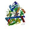


 PDBj
PDBj

 Assembly


 Mass: 129.164 Da / Num. of mol.: 1 / Source method: obtained synthetically / Formula: C4H11N5 / Comment: medication*YM
 Mass: 616.487 Da / Num. of mol.: 1 / Source method: obtained synthetically / Formula: C34H32FeN4O4
 Mass: 18.015 Da / Num. of mol.: 26 / Source method: isolated from a natural source / Formula: H2O
 Sample preparation
 / Beamline: BL7-1 / Wavelength: 1
  Processing