 Movie
Movie Controller


+ Open data

- Basic information
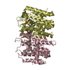
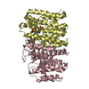
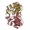
| Entry | Database: PDB / ID: 5fby | ||||||
|---|---|---|---|---|---|---|---|
| Title | Crystal structure of ctSPD | ||||||
 Components Components |
| ||||||
 Keywords Keywords |  HYDROLASE / cohesin / complex HYDROLASE / cohesin / complex | ||||||
| Function / homology |  Function and homology information Function and homology informationnuclear chromosome segregation / separase / nuclear division / cysteine-type endopeptidase activity / proteolysis / nucleusSimilarity search - Function | ||||||
| Biological species |  Chaetomium thermophilum (fungus) Chaetomium thermophilum (fungus)synthetic construct (others) | ||||||
| Method | X-RAY DIFFRACTION / SYNCHROTRON / Resolution: 1.898 Å | ||||||
 Authors Authors | Lin, Z. / Luo, X. / Yu, H. | ||||||
 Citation Citation | Journal: Nature / Year: 2016 Title: Structural basis of cohesin cleavage by separase. Authors: Lin, Z. / Luo, X. / Yu, H. | ||||||
| History |
|
- Structure visualization
| Structure viewer | Molecule: MolmilJmol/JSmol |
|---|
- Downloads & links
-Download
| PDBx/mmCIF format | 5fby.cif.gz | 218.3 KB | Display | PDBx/mmCIF format |
|---|---|---|---|---|
| PDB format | pdb5fby.ent.gz | 171.9 KB | Display | PDB format |
| PDBx/mmJSON format | 5fby.json.gz | Tree view | PDBx/mmJSON format | |
| Others |  Other downloads Other downloads |
-Validation report
| Arichive directory | https://data.pdbj.org/pub/pdb/validation_reports/fb/5fbyftp://data.pdbj.org/pub/pdb/validation_reports/fb/5fby | HTTPS FTP |
|---|
-Related structure data


-Links
 PDBj
PDBj- Assembly
| Deposited unit | 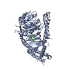
| ||||||||
|---|---|---|---|---|---|---|---|---|---|
| 1 |
| ||||||||
| Unit cell |
|
-Components
| #1: Protein | Mass: 62462.699 Da / Num. of mol.: 1 / Fragment: unp residues 1621-2223 Source method: isolated from a genetically manipulated source Source: (gene. exp.) Chaetomium thermophilum (strain DSM 1495 / CBS 144.50 / IMI 039719) (fungus)Strain: DSM 1495 / CBS 144.50 / IMI 039719 / Gene: CTHT_0070540 / Production host:  Escherichia coli (E. coli) / References: UniProt: G0SHM3 Escherichia coli (E. coli) / References: UniProt: G0SHM3 |
|---|---|
| #2: Protein/peptide | Mass: 2877.183 Da / Num. of mol.: 1 Source method: isolated from a genetically manipulated source Source: (gene. exp.) synthetic construct (others) / Production host: Escherichia coli (E. coli) |
| #3: Water | ChemComp-HOH / Water Mass: 18.015 Da / Num. of mol.: 394 / Source method: isolated from a natural source / Formula: H2O Mass: 18.015 Da / Num. of mol.: 394 / Source method: isolated from a natural source / Formula: H2O |
-Experimental details
-Experiment
| Experiment | Method: X-RAY DIFFRACTION |
|---|
- Sample preparation
| Crystal | Density Matthews: 2.27 Å3/Da / Density % sol: 45.7 % |
|---|---|
| Crystal grow | Temperature: 293 K / Method: vapor diffusion, hanging drop / Details: PEG3350, ammonium citrate tribasic |
-Data collection
| Diffraction | Mean temperature: 100 K |
|---|---|
| Diffraction source | Source: SYNCHROTRON / Site: ALS  / Beamline: 8.2.1 / Wavelength: 0.979 Å / Beamline: 8.2.1 / Wavelength: 0.979 Å |
| Detector | Type: ADSC QUANTUM 315r / Detector: CCD / Date: Jan 27, 2015 |
| Radiation | Protocol: SINGLE WAVELENGTH / Monochromatic (M) / Laue (L): M / Scattering type: x-ray |
| Radiation wavelength | Wavelength: 0.979 Å / Relative weight: 1 |
| Reflection | Resolution: 1.898→50 Å / Num. obs: 47718 / % possible obs: 100 % / Redundancy: 7.2 % / Net I/σ(I): 27.7 |
- Processing
| Software |
| |||||||||||||||||||||||||||||||||||||||||||||||||||||||||||||||||||||||||||||||||||||||||||||||||||||||||||||||||||||||||||||||||||||||||||||||||||||||||||||||||||||||||||||||
|---|---|---|---|---|---|---|---|---|---|---|---|---|---|---|---|---|---|---|---|---|---|---|---|---|---|---|---|---|---|---|---|---|---|---|---|---|---|---|---|---|---|---|---|---|---|---|---|---|---|---|---|---|---|---|---|---|---|---|---|---|---|---|---|---|---|---|---|---|---|---|---|---|---|---|---|---|---|---|---|---|---|---|---|---|---|---|---|---|---|---|---|---|---|---|---|---|---|---|---|---|---|---|---|---|---|---|---|---|---|---|---|---|---|---|---|---|---|---|---|---|---|---|---|---|---|---|---|---|---|---|---|---|---|---|---|---|---|---|---|---|---|---|---|---|---|---|---|---|---|---|---|---|---|---|---|---|---|---|---|---|---|---|---|---|---|---|---|---|---|---|---|---|---|---|---|---|
| Refinement | Resolution: 1.898→49.381 Å / SU ML: 0.17 / Cross valid method: FREE R-VALUE / σ(F): 1.35 / Phase error: 18.62 / Stereochemistry target values: ML
| |||||||||||||||||||||||||||||||||||||||||||||||||||||||||||||||||||||||||||||||||||||||||||||||||||||||||||||||||||||||||||||||||||||||||||||||||||||||||||||||||||||||||||||||
| Solvent computation | Shrinkage radii: 0.9 Å / VDW probe radii: 1.11 Å / Solvent model: FLAT BULK SOLVENT MODEL | |||||||||||||||||||||||||||||||||||||||||||||||||||||||||||||||||||||||||||||||||||||||||||||||||||||||||||||||||||||||||||||||||||||||||||||||||||||||||||||||||||||||||||||||
| Refinement step | Cycle: LAST / Resolution: 1.898→49.381 Å
| |||||||||||||||||||||||||||||||||||||||||||||||||||||||||||||||||||||||||||||||||||||||||||||||||||||||||||||||||||||||||||||||||||||||||||||||||||||||||||||||||||||||||||||||
| Refine LS restraints |
| |||||||||||||||||||||||||||||||||||||||||||||||||||||||||||||||||||||||||||||||||||||||||||||||||||||||||||||||||||||||||||||||||||||||||||||||||||||||||||||||||||||||||||||||
| LS refinement shell |
| |||||||||||||||||||||||||||||||||||||||||||||||||||||||||||||||||||||||||||||||||||||||||||||||||||||||||||||||||||||||||||||||||||||||||||||||||||||||||||||||||||||||||||||||
| Refinement TLS params. | Method: refined / Refine-ID: X-RAY DIFFRACTION
| |||||||||||||||||||||||||||||||||||||||||||||||||||||||||||||||||||||||||||||||||||||||||||||||||||||||||||||||||||||||||||||||||||||||||||||||||||||||||||||||||||||||||||||||
| Refinement TLS group |
|
