 Movie
Movie Controller

[English] 日本語
 Yorodumi
- PDB-5er8: Crystal structure of cyclization domain of Phomopsis amygdali fus... -
+ Open data

- Basic information
| Entry | Database: PDB / ID: 5er8 | ||||||
|---|---|---|---|---|---|---|---|
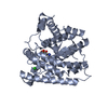
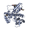
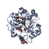
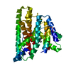
| Title | Crystal structure of cyclization domain of Phomopsis amygdali fusicoccadiene synthase complexed with manganese ions and neridronate | ||||||
 Components Components | Fusicoccadiene synthase Fusicocca-2,10(14)-diene synthase Fusicocca-2,10(14)-diene synthase | ||||||
 Keywords Keywords | LYASE / diterpene cyclase / terpenoids | ||||||
| Function / homology |  Function and homology informationfusicocca-2,10(14)-diene synthase / alcohol biosynthetic process / mycotoxin biosynthetic process / geranylgeranyl diphosphate synthase / farnesyltranstransferase activity / isoprenoid biosynthetic process / lyase activity / metal ion binding Function and homology informationfusicocca-2,10(14)-diene synthase / alcohol biosynthetic process / mycotoxin biosynthetic process / geranylgeranyl diphosphate synthase / farnesyltranstransferase activity / isoprenoid biosynthetic process / lyase activity / metal ion bindingSimilarity search - Function | ||||||
| Biological species |  Phomopsis amygdali (fungus) Phomopsis amygdali (fungus) | ||||||
| Method | X-RAY DIFFRACTION / SYNCHROTRON / SAD / Resolution: 2.5 Å | ||||||
 Authors Authors | Chen, M. / Christianson, D.W. | ||||||
 Citation Citation | Journal: Acs Chem.Biol. / Year: 2016 Title: Structure and Function of Fusicoccadiene Synthase, a Hexameric Bifunctional Diterpene Synthase. Authors: Chen, M. / Chou, W.K. / Toyomasu, T. / Cane, D.E. / Christianson, D.W. | ||||||
| History |
|
- Structure visualization
| Structure viewer | Molecule: MolmilJmol/JSmol |
|---|
- Downloads & links
-Download
| PDBx/mmCIF format | 5er8.cif.gz | 136.9 KB | Display | PDBx/mmCIF format |
|---|---|---|---|---|
| PDB format | pdb5er8.ent.gz | 110.4 KB | Display | PDB format |
| PDBx/mmJSON format | 5er8.json.gz | Tree view | PDBx/mmJSON format | |
| Others |  Other downloads Other downloads |
-Validation report
| Arichive directory | https://data.pdbj.org/pub/pdb/validation_reports/er/5er8ftp://data.pdbj.org/pub/pdb/validation_reports/er/5er8 | HTTPS FTP |
|---|
-Related structure data


-Links
 PDBj
PDBj
- Assembly
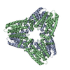
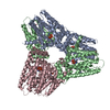
| Deposited unit | 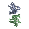
| ||||||||
|---|---|---|---|---|---|---|---|---|---|
| 1 | 
| ||||||||
| 2 | 
| ||||||||
| Unit cell |
|
-Components
| #1: Protein | Fusicocca-2,10(14)-diene synthase / FS / PaDC4:GGS / Diterpene cyclase 4 / DC 4 Mass: 41693.266 Da / Num. of mol.: 2 / Fragment: Fusicocca-2,10(14)-diene synthase, residues 1-344 Source method: isolated from a genetically manipulated source Details: Neridronate / Source: (gene. exp.) Phomopsis amygdali (fungus) / Gene: PaFS / Production host:  Escherichia coli (E. coli) Escherichia coli (E. coli)References: UniProt: A2PZA5, fusicocca-2,10(14)-diene synthase#2: Chemical | ChemComp-MN /   Mass: 54.938 Da / Num. of mol.: 9 / Source method: obtained synthetically / Formula: Mn Mass: 54.938 Da / Num. of mol.: 9 / Source method: obtained synthetically / Formula: Mn#3: Chemical | Neridronic acid  Mass: 277.149 Da / Num. of mol.: 2 / Source method: obtained synthetically / Formula: C6H17NO7P2 Mass: 277.149 Da / Num. of mol.: 2 / Source method: obtained synthetically / Formula: C6H17NO7P2#4: Chemical | Chloride  Mass: 35.453 Da / Num. of mol.: 2 / Source method: obtained synthetically / Formula: Cl Mass: 35.453 Da / Num. of mol.: 2 / Source method: obtained synthetically / Formula: Cl#5: Water | ChemComp-HOH / | Water Mass: 18.015 Da / Num. of mol.: 76 / Source method: isolated from a natural source / Formula: H2O Mass: 18.015 Da / Num. of mol.: 76 / Source method: isolated from a natural source / Formula: H2O |
|---|
-Experimental details
-Experiment
| Experiment | Method: X-RAY DIFFRACTION |
|---|
- Sample preparation
| Crystal | Density Matthews: 4.14 Å3/Da / Density % sol: 70.29 % |
|---|---|
| Crystal grow | Temperature: 277 K / Method: vapor diffusion, sitting drop Details: 1.05 M sodium formate, 0.075 M manganese formate, 0.1 M sodium acetate (pH 4.4) |
-Data collection
| Diffraction | Mean temperature: 100 K |
|---|---|
| Diffraction source | Source: SYNCHROTRON / Site: SSRL  / Beamline: BL14-1 / Wavelength: 1.18 Å / Beamline: BL14-1 / Wavelength: 1.18 Å |
| Detector | Type: MARMOSAIC 325 mm CCD / Detector: CCD / Date: Jun 4, 2015 |
| Radiation | Protocol: SINGLE WAVELENGTH / Monochromatic (M) / Laue (L): M / Scattering type: x-ray |
| Radiation wavelength | Wavelength: 1.18 Å / Relative weight: 1 |
| Reflection | Resolution: 2.5→43.4 Å / Num. obs: 48128 / % possible obs: 100 % / Redundancy: 8.4 % / Net I/σ(I): 30 |
- Processing
| Software |
| ||||||||||||||||||||||||||||||||||||||||||||||||||||||||||||||||||||||||||||||||||||||||||||||||||||||||||||||||||||||||||||||
|---|---|---|---|---|---|---|---|---|---|---|---|---|---|---|---|---|---|---|---|---|---|---|---|---|---|---|---|---|---|---|---|---|---|---|---|---|---|---|---|---|---|---|---|---|---|---|---|---|---|---|---|---|---|---|---|---|---|---|---|---|---|---|---|---|---|---|---|---|---|---|---|---|---|---|---|---|---|---|---|---|---|---|---|---|---|---|---|---|---|---|---|---|---|---|---|---|---|---|---|---|---|---|---|---|---|---|---|---|---|---|---|---|---|---|---|---|---|---|---|---|---|---|---|---|---|---|---|
| Refinement | Method to determine structure: SAD / Resolution: 2.5→43.386 Å / Cross valid method: FREE R-VALUE / σ(F): 1.34 / Phase error: 37.26 / Stereochemistry target values: TWIN_LSQ_F
| ||||||||||||||||||||||||||||||||||||||||||||||||||||||||||||||||||||||||||||||||||||||||||||||||||||||||||||||||||||||||||||||
| Solvent computation | Shrinkage radii: 0.9 Å / VDW probe radii: 1.11 Å / Solvent model: FLAT BULK SOLVENT MODEL | ||||||||||||||||||||||||||||||||||||||||||||||||||||||||||||||||||||||||||||||||||||||||||||||||||||||||||||||||||||||||||||||
| Refinement step | Cycle: LAST / Resolution: 2.5→43.386 Å
| ||||||||||||||||||||||||||||||||||||||||||||||||||||||||||||||||||||||||||||||||||||||||||||||||||||||||||||||||||||||||||||||
| Refine LS restraints |
| ||||||||||||||||||||||||||||||||||||||||||||||||||||||||||||||||||||||||||||||||||||||||||||||||||||||||||||||||||||||||||||||
| LS refinement shell |
|
