 Movie
Movie Controller

[English] 日本語
 Yorodumi
- PDB-5dgs: Crystal structure of human FPPS in complex with the monophosphona... -
+ Open data

- Basic information
| Entry | Database: PDB / ID: 5dgs | ||||||
|---|---|---|---|---|---|---|---|
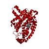
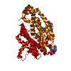
| Title | Crystal structure of human FPPS in complex with the monophosphonate compound 15 | ||||||
 Components Components | Farnesyl pyrophosphate synthase Dimethylallyltranstransferase Dimethylallyltranstransferase | ||||||
 Keywords Keywords | TRANSFERASE / ISOPRENE BIOSYNTHESIS / CHOLESTEROL BIOSYNTHESIS | ||||||
| Function / homology |  Function and homology information Function and homology informationgeranyl diphosphate biosynthetic process / dimethylallyltranstransferase / (2E,6E)-farnesyl diphosphate synthase / Cholesterol biosynthesis / farnesyl diphosphate biosynthetic process / dimethylallyltranstransferase activity / geranyltranstransferase activity / cholesterol biosynthetic process / Activation of gene expression by SREBF (SREBP) / RNA binding ...geranyl diphosphate biosynthetic process / dimethylallyltranstransferase / (2E,6E)-farnesyl diphosphate synthase / Cholesterol biosynthesis / farnesyl diphosphate biosynthetic process / dimethylallyltranstransferase activity / geranyltranstransferase activity / cholesterol biosynthetic process / Activation of gene expression by SREBF (SREBP) / RNA binding / nucleoplasm / metal ion binding / cytosol / cytoplasmSimilarity search - Function | ||||||
| Biological species |  Homo sapiens (human) Homo sapiens (human) | ||||||
| Method | X-RAY DIFFRACTION / FOURIER SYNTHESIS / Resolution: 2.62 Å | ||||||
 Authors Authors | Rondeau, J.M. / Bourgier, E. / Lehmann, S. | ||||||
 Citation Citation | Journal: Angew.Chem.Int.Ed.Engl. / Year: 2015 Title: A General Strategy for Targeting Drugs to Bone. Authors: Jahnke, W. / Bold, G. / Marzinzik, A.L. / Ofner, S. / Pelle, X. / Cotesta, S. / Bourgier, E. / Lehmann, S. / Henry, C. / Hemmig, R. / Stauffer, F. / Hartwieg, J.C. / Green, J.R. / Rondeau, J.M. #1: Journal: Nat.Chem.Biol. / Year: 2010Title: Allosteric non-bisphosphonate FPPS inhibitors identified by fragment-based discovery. Authors: Jahnke, W. / Rondeau, J.M. / Cotesta, S. / Marzinzik, A. / Pelle, X. / Geiser, M. / Strauss, A. / Gotte, M. / Bitsch, F. / Hemmig, R. / Henry, C. / Lehmann, S. / Glickman, J.F. / Roddy, T.P. ...Authors: Jahnke, W. / Rondeau, J.M. / Cotesta, S. / Marzinzik, A. / Pelle, X. / Geiser, M. / Strauss, A. / Gotte, M. / Bitsch, F. / Hemmig, R. / Henry, C. / Lehmann, S. / Glickman, J.F. / Roddy, T.P. / Stout, S.J. / Green, J.R. #2: Journal: Chemmedchem / Year: 2015Title: Discovery of Novel Allosteric Non-Bisphosphonate Inhibitors of Farnesyl Pyrophosphate Synthase by Integrated Lead Finding. Authors: Marzinzik, A.L. / Amstutz, R. / Bold, G. / Bourgier, E. / Cotesta, S. / Glickman, J.F. / Gotte, M. / Henry, C. / Lehmann, S. / Hartwieg, J.C. / Ofner, S. / Pelle, X. / Roddy, T.P. / Rondeau, ...Authors: Marzinzik, A.L. / Amstutz, R. / Bold, G. / Bourgier, E. / Cotesta, S. / Glickman, J.F. / Gotte, M. / Henry, C. / Lehmann, S. / Hartwieg, J.C. / Ofner, S. / Pelle, X. / Roddy, T.P. / Rondeau, J.M. / Stauffer, F. / Stout, S.J. / Widmer, A. / Zimmermann, J. / Zoller, T. / Jahnke, W. | ||||||
| History |
|
- Structure visualization
| Structure viewer | Molecule: MolmilJmol/JSmol |
|---|
- Downloads & links
-Download
| PDBx/mmCIF format | 5dgs.cif.gz | 157.5 KB | Display | PDBx/mmCIF format |
|---|---|---|---|---|
| PDB format | pdb5dgs.ent.gz | 124.1 KB | Display | PDB format |
| PDBx/mmJSON format | 5dgs.json.gz | Tree view | PDBx/mmJSON format | |
| Others |  Other downloads Other downloads |
-Validation report
| Arichive directory | https://data.pdbj.org/pub/pdb/validation_reports/dg/5dgsftp://data.pdbj.org/pub/pdb/validation_reports/dg/5dgs | HTTPS FTP |
|---|
-Related structure data


-Links
 PDBj
PDBj

- Assembly
| Deposited unit | 
| ||||||||
|---|---|---|---|---|---|---|---|---|---|
| 1 | 
| ||||||||
| Unit cell |
|
-Components
| #1: Protein | Dimethylallyltranstransferase / FPS / (2E / 6E)-farnesyl diphosphate synthase / Dimethylallyltranstransferase / Farnesyl ...FPS / (2E / 6E)-farnesyl diphosphate synthase / Dimethylallyltranstransferase / Farnesyl diphosphate synthase / Geranyltranstransferase Mass: 40183.855 Da / Num. of mol.: 1 / Fragment: UNP residues 72-419 Source method: isolated from a genetically manipulated source Source: (gene. exp.) Homo sapiens (human) / Gene: FDPS, FPS, KIAA1293 / Plasmid: pET28 / Production host:  Escherichia coli BL21(DE3) (bacteria) / Variant (production host): tuner Escherichia coli BL21(DE3) (bacteria) / Variant (production host): tunerReferences: UniProt: P14324, (2E,6E)-farnesyl diphosphate synthase, dimethylallyltranstransferase |
|---|---|
| #2: Chemical | ChemComp-5A7 / {(  Mass: 418.382 Da / Num. of mol.: 1 / Source method: obtained synthetically / Formula: C23H19N2O4P Mass: 418.382 Da / Num. of mol.: 1 / Source method: obtained synthetically / Formula: C23H19N2O4P |
| #3: Water | ChemComp-HOH / Water Mass: 18.015 Da / Num. of mol.: 48 / Source method: isolated from a natural source / Formula: H2O Mass: 18.015 Da / Num. of mol.: 48 / Source method: isolated from a natural source / Formula: H2O |
-Experimental details
-Experiment
| Experiment | Method: X-RAY DIFFRACTION / Number of used crystals: 1 |
|---|
- Sample preparation
| Crystal | Density Matthews: 2.86 Å3/Da / Density % sol: 57.05 % |
|---|---|
| Crystal grow | Temperature: 292 K / Method: vapor diffusion, hanging drop / pH: 4.7 / Details: 1.2M sodium potassium phosphate, 25% glycerol |
-Data collection
| Diffraction | Mean temperature: 100 K |
|---|---|
| Diffraction source | Source: ROTATING ANODE / Type: RIGAKU FR-E SUPERBRIGHT / Wavelength: 1.54178 Å |
| Detector | Type: RIGAKU SATURN 92 / Detector: CCD / Date: Apr 18, 2007 |
| Radiation | Monochromator: MIRRORS / Protocol: SINGLE WAVELENGTH / Monochromatic (M) / Laue (L): M / Scattering type: x-ray |
| Radiation wavelength | Wavelength: 1.54178 Å / Relative weight: 1 |
| Reflection | Resolution: 2.62→78.46 Å / Num. obs: 14536 / % possible obs: 99.9 % / Observed criterion σ(I): -3 / Redundancy: 13.8 % / Rmerge(I) obs: 0.117 / Net I/σ(I): 17.84 |
| Reflection shell | Resolution: 2.62→2.72 Å / Redundancy: 13.8 % / Rmerge(I) obs: 0.537 / Mean I/σ(I) obs: 5.57 / % possible all: 99.9 |
- Processing
| Software |
| ||||||||||||||||||||||||||||||||||||||||||||||||||||||||||||||||||||||||||||||||||||||||||||||||||||||||||||
|---|---|---|---|---|---|---|---|---|---|---|---|---|---|---|---|---|---|---|---|---|---|---|---|---|---|---|---|---|---|---|---|---|---|---|---|---|---|---|---|---|---|---|---|---|---|---|---|---|---|---|---|---|---|---|---|---|---|---|---|---|---|---|---|---|---|---|---|---|---|---|---|---|---|---|---|---|---|---|---|---|---|---|---|---|---|---|---|---|---|---|---|---|---|---|---|---|---|---|---|---|---|---|---|---|---|---|---|---|---|
| Refinement | Method to determine structure: FOURIER SYNTHESIS / Resolution: 2.62→23.17 Å / Cor.coef. Fo:Fc: 0.9479 / Cor.coef. Fo:Fc free: 0.912 / SU R Cruickshank DPI: 0.468 / Cross valid method: THROUGHOUT / σ(F): 0 / SU R Blow DPI: 0.494 / SU Rfree Blow DPI: 0.281 / SU Rfree Cruickshank DPI: 0.282
| ||||||||||||||||||||||||||||||||||||||||||||||||||||||||||||||||||||||||||||||||||||||||||||||||||||||||||||
| Displacement parameters | Biso max: 176.62 Å2 / Biso mean: 70.32 Å2 / Biso min: 19.21 Å2
| ||||||||||||||||||||||||||||||||||||||||||||||||||||||||||||||||||||||||||||||||||||||||||||||||||||||||||||
| Refine analyze | Luzzati coordinate error free: 0.385 Å | ||||||||||||||||||||||||||||||||||||||||||||||||||||||||||||||||||||||||||||||||||||||||||||||||||||||||||||
| Refinement step | Cycle: final / Resolution: 2.62→23.17 Å
| ||||||||||||||||||||||||||||||||||||||||||||||||||||||||||||||||||||||||||||||||||||||||||||||||||||||||||||
| Refine LS restraints |
| ||||||||||||||||||||||||||||||||||||||||||||||||||||||||||||||||||||||||||||||||||||||||||||||||||||||||||||
| LS refinement shell | Resolution: 2.62→2.83 Å / Total num. of bins used: 7
| ||||||||||||||||||||||||||||||||||||||||||||||||||||||||||||||||||||||||||||||||||||||||||||||||||||||||||||
| Refinement TLS params. | Method: refined / Details: FPPS / Origin x: 10.1454 Å / Origin y: 79.8974 Å / Origin z: 26.6041 Å
| ||||||||||||||||||||||||||||||||||||||||||||||||||||||||||||||||||||||||||||||||||||||||||||||||||||||||||||
| Refinement TLS group | Selection: all / Selection details: { F|* } |
