 Movie
Movie Controller


+ Open data

- Basic information
| Entry | Database: PDB / ID: 5ctq | ||||||
|---|---|---|---|---|---|---|---|
| Title | Crystal structure of human SART3/TIP110 half-a TPR (HAT) domain | ||||||
 Components Components | Squamous cell carcinoma antigen recognized by T-cells 3 | ||||||
 Keywords Keywords |  IMMUNE SYSTEM / NUCLEAR PROTEIN / RNA BINDING PROTEIN / half a tetratricopeptide repeat (HAT) domain / Protein transporter IMMUNE SYSTEM / NUCLEAR PROTEIN / RNA BINDING PROTEIN / half a tetratricopeptide repeat (HAT) domain / Protein transporter | ||||||
| Function / homology |  Function and homology information Function and homology informationU6atac snRNA binding / ASAP complex / U4 snRNA binding / hematopoietic stem cell proliferation / transcription elongation-coupled chromatin remodeling / ubiquitin-specific protease binding / homeostasis of number of cells / spliceosomal tri-snRNP complex assembly / Cajal body / U6 snRNA binding ...U6atac snRNA binding / ASAP complex / U4 snRNA binding / hematopoietic stem cell proliferation / transcription elongation-coupled chromatin remodeling / ubiquitin-specific protease binding / homeostasis of number of cells / spliceosomal tri-snRNP complex assembly / Cajal body / U6 snRNA binding / spliceosomal snRNP assembly / cell morphogenesis / mRNA splicing, via spliceosome / nucleosome assembly / histone binding / regulation of gene expression / nuclear speck / RNA binding / nucleoplasm / nucleus / cytoplasmSimilarity search - Function | ||||||
| Biological species |  Homo sapiens (human) Homo sapiens (human) | ||||||
| Method | X-RAY DIFFRACTION / SYNCHROTRON / SAD / Resolution: 2.6 Å | ||||||
 Authors Authors | Park, J.K. / Kim, E.E. | ||||||
| Funding support | Korea, Democratic People's Republic Of, 1items
| ||||||
 Citation Citation | Journal: Nucleic Acids Res. / Year: 2016 Title: Structural basis for recruiting and shuttling of the spliceosomal deubiquitinase USP4 by SART3 Authors: Park, J.K. / Das, T. / Song, E.J. / Kim, E.E. | ||||||
| History |
|
- Structure visualization
| Structure viewer | Molecule: MolmilJmol/JSmol |
|---|
- Downloads & links
-Download
| PDBx/mmCIF format | 5ctq.cif.gz | 416.9 KB | Display | PDBx/mmCIF format |
|---|---|---|---|---|
| PDB format | pdb5ctq.ent.gz | 341.1 KB | Display | PDB format |
| PDBx/mmJSON format | 5ctq.json.gz | Tree view | PDBx/mmJSON format | |
| Others |  Other downloads Other downloads |
-Validation report
| Arichive directory | https://data.pdbj.org/pub/pdb/validation_reports/ct/5ctqftp://data.pdbj.org/pub/pdb/validation_reports/ct/5ctq | HTTPS FTP |
|---|
-Related structure data


-Links
 PDBj
PDBj
- Assembly
| Deposited unit | 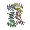
| ||||||||
|---|---|---|---|---|---|---|---|---|---|
| 1 | 
| ||||||||
| 2 | 
| ||||||||
| Unit cell |
|
-Components
| #1: Protein | Mass: 65820.547 Da / Num. of mol.: 4 / Fragment: UNP residues 94-611 Source method: isolated from a genetically manipulated source Source: (gene. exp.) Homo sapiens (human) / Gene: SART3, KIAA0156, TIP110 / Production host:  Escherichia coli (E. coli) / References: UniProt: Q15020 Escherichia coli (E. coli) / References: UniProt: Q15020#2: Water | ChemComp-HOH / | Water Mass: 18.015 Da / Num. of mol.: 281 / Source method: isolated from a natural source / Formula: H2O Mass: 18.015 Da / Num. of mol.: 281 / Source method: isolated from a natural source / Formula: H2O |
|---|
-Experimental details
-Experiment
| Experiment | Method: X-RAY DIFFRACTION |
|---|
- Sample preparation
| Crystal | Density Matthews: 2.88 Å3/Da / Density % sol: 57.26 % |
|---|---|
| Crystal grow | Temperature: 293 K / Method: vapor diffusion, hanging drop / Details: 200mM sodium formate pH 7.5, 30% PEG4000 |
-Data collection
| Diffraction | Mean temperature: 100 K |
|---|---|
| Diffraction source | Source: SYNCHROTRON / Site: PAL/PLS  / Beamline: 5C (4A) / Wavelength: 0.9794 Å / Beamline: 5C (4A) / Wavelength: 0.9794 Å |
| Detector | Type: ADSC QUANTUM 315r / Detector: CCD / Date: May 28, 2012 |
| Radiation | Protocol: SINGLE WAVELENGTH / Monochromatic (M) / Laue (L): M / Scattering type: x-ray |
| Radiation wavelength | Wavelength: 0.9794 Å / Relative weight: 1 |
| Reflection | Resolution: 2.6→50 Å / Num. obs: 87186 / % possible obs: 95.1 % / Redundancy: 2.5 % / Rmerge(I) obs: 0.097 / Net I/σ(I): 14.5 |
| Reflection shell | Resolution: 2.6→2.69 Å / Redundancy: 1.6 % / Rmerge(I) obs: 0.395 / Mean I/σ(I) obs: 2.07 / % possible all: 82.9 |
- Processing
| Software |
| |||||||||||||||||||||||||||||||||||||||||||||||||||||||||||||||||||||||||||||||||||||||||||||||||||||||||
|---|---|---|---|---|---|---|---|---|---|---|---|---|---|---|---|---|---|---|---|---|---|---|---|---|---|---|---|---|---|---|---|---|---|---|---|---|---|---|---|---|---|---|---|---|---|---|---|---|---|---|---|---|---|---|---|---|---|---|---|---|---|---|---|---|---|---|---|---|---|---|---|---|---|---|---|---|---|---|---|---|---|---|---|---|---|---|---|---|---|---|---|---|---|---|---|---|---|---|---|---|---|---|---|---|---|---|
| Refinement | Method to determine structure: SAD / Resolution: 2.6→38.15 Å / SU ML: 0.49 / Cross valid method: FREE R-VALUE / σ(F): 1.47 / Phase error: 31.97 / Stereochemistry target values: ML
| |||||||||||||||||||||||||||||||||||||||||||||||||||||||||||||||||||||||||||||||||||||||||||||||||||||||||
| Solvent computation | Shrinkage radii: 0.73 Å / VDW probe radii: 1 Å / Solvent model: FLAT BULK SOLVENT MODEL / Bsol: 14.321 Å2 / ksol: 0.268 e/Å3 | |||||||||||||||||||||||||||||||||||||||||||||||||||||||||||||||||||||||||||||||||||||||||||||||||||||||||
| Displacement parameters |
| |||||||||||||||||||||||||||||||||||||||||||||||||||||||||||||||||||||||||||||||||||||||||||||||||||||||||
| Refinement step | Cycle: LAST / Resolution: 2.6→38.15 Å
| |||||||||||||||||||||||||||||||||||||||||||||||||||||||||||||||||||||||||||||||||||||||||||||||||||||||||
| Refine LS restraints |
| |||||||||||||||||||||||||||||||||||||||||||||||||||||||||||||||||||||||||||||||||||||||||||||||||||||||||
| LS refinement shell |
|
