 Movie
Movie Controller

[English] 日本語
 Yorodumi
- PDB-5but: Crystal structure of inactive conformation of KtrAB K+ transporter -
+ Open data

- Basic information
| Entry | Database: PDB / ID: 5but | ||||||
|---|---|---|---|---|---|---|---|
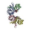
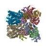
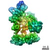
| Title | Crystal structure of inactive conformation of KtrAB K+ transporter | ||||||
 Components Components |
| ||||||
 Keywords Keywords |  MEMBRANE PROTEIN / membrane protein complex MEMBRANE PROTEIN / membrane protein complex | ||||||
| Function / homology |  Function and homology information Function and homology informationpotassium:chloride symporter activity / monoatomic cation transmembrane transporter activity / potassium ion transport / identical protein binding / plasma membraneSimilarity search - Function | ||||||
| Biological species |  Bacillus subtilis (bacteria) Bacillus subtilis (bacteria) | ||||||
| Method | X-RAY DIFFRACTION / SYNCHROTRON / MOLECULAR REPLACEMENT / Resolution: 5.97 Å | ||||||
 Authors Authors | Vieira-Pires, R.S. / Morais-Cabral, J.H. | ||||||
| Funding support |  Portugal, 1items Portugal, 1items
| ||||||
 Citation Citation | Journal: Plos Biol. / Year: 2016 Title: Dissecting the Molecular Mechanism of Nucleotide-Dependent Activation of the KtrAB K+ Transporter. Authors: Szollosi, A. / Vieira-Pires, R.S. / Teixeira-Duarte, C.M. / Rocha, R. / Morais-Cabral, J.H. | ||||||
| History |
|
- Structure visualization
| Structure viewer | Molecule: MolmilJmol/JSmol |
|---|
- Downloads & links
-Download
| PDBx/mmCIF format | 5but.cif.gz | 457.9 KB | Display | PDBx/mmCIF format |
|---|---|---|---|---|
| PDB format | pdb5but.ent.gz | 364.1 KB | Display | PDB format |
| PDBx/mmJSON format | 5but.json.gz | Tree view | PDBx/mmJSON format | |
| Others |  Other downloads Other downloads |
-Validation report
| Arichive directory | https://data.pdbj.org/pub/pdb/validation_reports/bu/5butftp://data.pdbj.org/pub/pdb/validation_reports/bu/5but | HTTPS FTP |
|---|
-Related structure data


-Links
 PDBj
PDBj
- Assembly
| Deposited unit | 
| ||||||||
|---|---|---|---|---|---|---|---|---|---|
| 1 | 
| ||||||||
| 2 | 
| ||||||||
| Unit cell |
|
-Components
| #1: Protein | Mass: 32038.717 Da / Num. of mol.: 4 / Fragment: regulatory domain,regulatory domain Mutation: truncation of C-terminal domain,truncation of C-terminal domain Source method: isolated from a genetically manipulated source Details: KtrA delta C is a fusion of two N-terminal domains of KtrA through the linker LEGS. It also includes a C to V mutation. Source: (gene. exp.) Bacillus subtilis (bacteria) / Gene: ktrA, yuaA, BSU31090 / Plasmid: PET24 / Production host: Escherichia coli BL21(DE3) (bacteria) / References: UniProt: O32080#2: Protein | Mass: 48158.039 Da / Num. of mol.: 4 / Fragment: membrane protein Source method: isolated from a genetically manipulated source Source: (gene. exp.) Bacillus subtilis (bacteria) / Gene: ktrB, yubG, BSU31100 / Plasmid: PET24 / Production host: Escherichia coli BL21(DE3) (bacteria) / References: UniProt: O32081#3: Chemical | ChemComp-K /   Mass: 39.098 Da / Num. of mol.: 4 / Source method: obtained synthetically / Formula: K Mass: 39.098 Da / Num. of mol.: 4 / Source method: obtained synthetically / Formula: K |
|---|
-Experimental details
-Experiment
| Experiment | Method: X-RAY DIFFRACTION / Number of used crystals: 1 |
|---|
- Sample preparation
| Crystal | Density Matthews: 3.86 Å3/Da / Density % sol: 68.12 % |
|---|---|
| Crystal grow | Temperature: 293 K / Method: vapor diffusion, sitting drop / pH: 8.5 / Details: 100 mM Tris-HCl, 16-22% PEG 400, 100-200 mM CaCl2 |
-Data collection
| Diffraction | Mean temperature: 100 K | ||||||||||||||||||||||||||||||||||||||||||||||||||||||||||||||||||||||||||||||||||||||||||||||||||||||||||||||
|---|---|---|---|---|---|---|---|---|---|---|---|---|---|---|---|---|---|---|---|---|---|---|---|---|---|---|---|---|---|---|---|---|---|---|---|---|---|---|---|---|---|---|---|---|---|---|---|---|---|---|---|---|---|---|---|---|---|---|---|---|---|---|---|---|---|---|---|---|---|---|---|---|---|---|---|---|---|---|---|---|---|---|---|---|---|---|---|---|---|---|---|---|---|---|---|---|---|---|---|---|---|---|---|---|---|---|---|---|---|---|---|
| Diffraction source | Source: SYNCHROTRON / Site: SOLEIL  / Beamline: PROXIMA 2 / Wavelength: 0.976 Å / Beamline: PROXIMA 2 / Wavelength: 0.976 Å | ||||||||||||||||||||||||||||||||||||||||||||||||||||||||||||||||||||||||||||||||||||||||||||||||||||||||||||||
| Detector | Type: ADSC QUANTUM 315r / Detector: CCD / Date: Mar 20, 2014 | ||||||||||||||||||||||||||||||||||||||||||||||||||||||||||||||||||||||||||||||||||||||||||||||||||||||||||||||
| Radiation | Protocol: SINGLE WAVELENGTH / Monochromatic (M) / Laue (L): M / Scattering type: x-ray | ||||||||||||||||||||||||||||||||||||||||||||||||||||||||||||||||||||||||||||||||||||||||||||||||||||||||||||||
| Radiation wavelength | Wavelength: 0.976 Å / Relative weight: 1 | ||||||||||||||||||||||||||||||||||||||||||||||||||||||||||||||||||||||||||||||||||||||||||||||||||||||||||||||
| Reflection | Resolution: 5.97→200 Å / Num. all: 12628 / Num. obs: 12628 / % possible obs: 98.2 % / Redundancy: 3.4 % / Rpim(I) all: 0.044 / Rrim(I) all: 0.083 / Rsym value: 0.07 / Net I/av σ(I): 8.56 / Net I/σ(I): 10.2 / Num. measured all: 42979 | ||||||||||||||||||||||||||||||||||||||||||||||||||||||||||||||||||||||||||||||||||||||||||||||||||||||||||||||
| Reflection shell | Diffraction-ID: 1 / Rejects: 0
|
- Processing
| Software |
| |||||||||||||||||||||||||||||||||||||||||||||||||||||||||||||||||||||||||||||
|---|---|---|---|---|---|---|---|---|---|---|---|---|---|---|---|---|---|---|---|---|---|---|---|---|---|---|---|---|---|---|---|---|---|---|---|---|---|---|---|---|---|---|---|---|---|---|---|---|---|---|---|---|---|---|---|---|---|---|---|---|---|---|---|---|---|---|---|---|---|---|---|---|---|---|---|---|---|---|
| Refinement | Method to determine structure: MOLECULAR REPLACEMENT Starting model: KtrB from 4J7C and KtrA-deltaC from 2HMS Resolution: 5.97→200 Å / Cross valid method: FREE R-VALUE / σ(F): 0
| |||||||||||||||||||||||||||||||||||||||||||||||||||||||||||||||||||||||||||||
| Solvent computation | Bsol: 261.776 Å2 | |||||||||||||||||||||||||||||||||||||||||||||||||||||||||||||||||||||||||||||
| Displacement parameters | Biso max: 342.98 Å2 / Biso mean: 342.9982 Å2 / Biso min: 342.98 Å2
| |||||||||||||||||||||||||||||||||||||||||||||||||||||||||||||||||||||||||||||
| Refinement step | Cycle: final / Resolution: 5.97→200 Å
| |||||||||||||||||||||||||||||||||||||||||||||||||||||||||||||||||||||||||||||
| Refine LS restraints |
| |||||||||||||||||||||||||||||||||||||||||||||||||||||||||||||||||||||||||||||
| LS refinement shell | Refine-ID: X-RAY DIFFRACTION / Total num. of bins used: 10
| |||||||||||||||||||||||||||||||||||||||||||||||||||||||||||||||||||||||||||||
| Xplor file |
|
