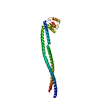
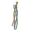
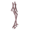
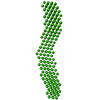
- PDB-5ayh: Structure of the entire dynein stalk region -
+
Open data
ID or keywords:
Loading...
-
Basic information
Entry
Database: PDB / ID: 5ayh
Title
Structure of the entire dynein stalk region
Components
Cytoplasmic dynein 1 heavy chain 1
Keywords
MOTOR PROTEIN / Microtubule
Function / homology
Function and homology information
COPI-independent Golgi-to-ER retrograde traffic / HSP90 chaperone cycle for steroid hormone receptors (SHR) in the presence of ligand / Amplification of signal from unattached kinetochores via a MAD2 inhibitory signal / COPI-mediated anterograde transport / cilium movement / Aggrephagy / positive regulation of intracellular transport / Mitotic Prometaphase / EML4 and NUDC in mitotic spindle formation / regulation of metaphase plate congression ...COPI-independent Golgi-to-ER retrograde traffic / HSP90 chaperone cycle for steroid hormone receptors (SHR) in the presence of ligand / Amplification of signal from unattached kinetochores via a MAD2 inhibitory signal / COPI-mediated anterograde transport / cilium movement / Aggrephagy / positive regulation of intracellular transport / Mitotic Prometaphase / EML4 and NUDC in mitotic spindle formation / regulation of metaphase plate congression / Resolution of Sister Chromatid Cohesion / establishment of spindle localization / positive regulation of spindle assembly / RHO GTPases Activate Formins / Separation of Sister Chromatids / Loss of Nlp from mitotic centrosomes / Recruitment of mitotic centrosome proteins and complexes / Loss of proteins required for interphase microtubule organization from the centrosome / Recruitment of NuMA to mitotic centrosomes / Anchoring of the basal body to the plasma membrane / AURKA Activation by TPX2 / Regulation of PLK1 Activity at G2/M Transition / manchette / P-body assembly / dynein complex / MHC class II antigen presentation / minus-end-directed microtubule motor activity / cytoplasmic dynein complex / retrograde axonal transport / dynein light intermediate chain binding / nuclear migration / dynein intermediate chain binding / cytoplasmic microtubule / cytoplasmic microtubule organization / stress granule assembly / regulation of mitotic spindle organization / axon cytoplasm / Neutrophil degranulation / mitotic spindle organization / filopodium / nuclear envelope / positive regulation of cold-induced thermogenesis / cell cortex / microtubule / axon / cell division / centrosome / neuronal cell body / ATP hydrolysis activity / ATP binding / cytoplasm Similarity search - Function
Dynein heavy chain, AAA 5 extension domain / Dynein heavy chain AAA lid domain / Dynein heavy chain, C-terminal domain / Dynein heavy chain, C-terminal domain, barrel region / Dynein heavy chain C-terminal domain / P-loop containing dynein motor region / Dynein heavy chain, tail / Dynein heavy chain, N-terminal region 1 / Dynein heavy chain / Dynein heavy chain region D6 P-loop domain ...Dynein heavy chain, AAA 5 extension domain / Dynein heavy chain AAA lid domain / Dynein heavy chain, C-terminal domain / Dynein heavy chain, C-terminal domain, barrel region / Dynein heavy chain C-terminal domain / P-loop containing dynein motor region / Dynein heavy chain, tail / Dynein heavy chain, N-terminal region 1 / Dynein heavy chain / Dynein heavy chain region D6 P-loop domain / Dynein heavy chain, linker / Dynein heavy chain, AAA module D4 / Dynein heavy chain, coiled coil stalk / Dynein heavy chain, hydrolytic ATP-binding dynein motor region / Dynein heavy chain, ATP-binding dynein motor region / Dynein heavy chain AAA lid domain / Dynein heavy chain AAA lid domain superfamily / Dynein heavy chain, domain 2, N-terminal / Dynein heavy chain, linker, subdomain 3 / Dynein heavy chain, AAA1 domain, small subdomain / Dynein heavy chain region D6 P-loop domain / Dynein heavy chain, N-terminal region 2 / Hydrolytic ATP binding site of dynein motor region / Microtubule-binding stalk of dynein motor / P-loop containing dynein motor region D4 / ATP-binding dynein motor region / Dynein heavy chain AAA lid domain / ATPases associated with a variety of cellular activities / AAA+ ATPase domain / P-loop containing nucleoside triphosphate hydrolase Similarity search - Domain/homology
In the structure databanks used in Yorodumi, some data are registered as the other names, "COVID-19 virus" and "2019-nCoV". Here are the details of the virus and the list of structure data.
Jan 31, 2019. EMDB accession codes are about to change! (news from PDBe EMDB page)
EMDB accession codes are about to change! (news from PDBe EMDB page)
The allocation of 4 digits for EMDB accession codes will soon come to an end. Whilst these codes will remain in use, new EMDB accession codes will include an additional digit and will expand incrementally as the available range of codes is exhausted. The current 4-digit format prefixed with “EMD-” (i.e. EMD-XXXX) will advance to a 5-digit format (i.e. EMD-XXXXX), and so on. It is currently estimated that the 4-digit codes will be depleted around Spring 2019, at which point the 5-digit format will come into force.
The EM Navigator/Yorodumi systems omit the EMD- prefix.
Related info.:Q: What is EMD? / ID/Accession-code notation in Yorodumi/EM Navigator
Yorodumi is a browser for structure data from EMDB, PDB, SASBDB, etc.
This page is also the successor to EM Navigator detail page, and also detail information page/front-end page for Omokage search.
The word "yorodu" (or yorozu) is an old Japanese word meaning "ten thousand". "mi" (miru) is to see.
Related info.:EMDB / PDB / SASBDB / Comparison of 3 databanks / Yorodumi Search / Aug 31, 2016. New EM Navigator & Yorodumi / Yorodumi Papers / Jmol/JSmol / Function and homology information / Changes in new EM Navigator and Yorodumi
 Movie
Movie Controller


 Open data
 Basic information
 Components
 Keywords
 MOTOR PROTEIN /
  Function and homology information
 Authors
 Japan, 1items
  Citation
 Structure visualization
 Downloads & links
 Other downloads
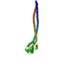


 PDBj
PDBj


 Assembly

 Sample preparation
 Processing