Entry Database : PDB / ID : 4zw2Title Crystal structure of the Mouse voltage gated calcium channel beta subunit isoform 1a in complex with Alpha Interaction Domain peptide. Voltage-dependent L-type calcium channel subunit alpha-1S Voltage-dependent L-type calcium channel subunit beta-1,Voltage-dependent L-type calcium channel subunit beta-1 Keywords / / / / Function / homology Function Domain/homology Component
/ / / / / / / / / / / / / / / / / / / / / / / / / / / / / / / / / / / / / / / / / / / / / / / / / / / / / / / / / / / / / / / / / / / / / / / / / / / / / / / / / / / / / / / / / / / / / / / Biological species Mus musculus (house mouse)Method / / Resolution : 1.86 Å Authors Norris, N.C. / Oakley, A.J. Journal : J. Biol. Chem. / Year : 2017Title : Structural and biophysical analyses of the skeletal dihydropyridine receptor beta subunit beta 1a reveal critical roles of domain interactions for stability.Authors : Norris, N.C. / Joseph, S. / Aditya, S. / Karunasekara, Y. / Board, P.G. / Dulhunty, A.F. / Oakley, A.J. / Casarotto, M.G. History Deposition May 19, 2015 Deposition site / Processing site Revision 1.0 Jun 1, 2016 Provider / Type Revision 1.1 Jul 27, 2016 Group Revision 1.2 Aug 2, 2017 Group / Derived calculationsCategory / citation_author / pdbx_struct_oper_listItem _citation.country / _citation.journal_abbrev ... _citation.country / _citation.journal_abbrev / _citation.journal_id_ASTM / _citation.journal_id_CSD / _citation.journal_id_ISSN / _citation.journal_volume / _citation.page_first / _citation.page_last / _citation.pdbx_database_id_DOI / _citation.pdbx_database_id_PubMed / _citation.title / _citation.year / _pdbx_struct_oper_list.symmetry_operation Revision 1.3 Sep 27, 2023 Group / Database references / Refinement descriptionCategory chem_comp_atom / chem_comp_bond ... chem_comp_atom / chem_comp_bond / database_2 / pdbx_initial_refinement_model Item / _database_2.pdbx_database_accession
Show all Show less
 Movie
Movie Controller

 Yorodumi
 Open data
 Basic information
 Components
 Keywords
 dihydropyridine receptor / CaVbeta / excitation contraction coupling / Alpha Interacting Domain
 Function and homology information
 Authors
 Citation
 Structure visualization
 Downloads & links
 Other downloads
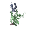


 PDBj
PDBj

 Assembly

 Mass: 150.173 Da / Num. of mol.: 2 / Source method: obtained synthetically / Formula: C6H14O4
 Mass: 18.015 Da / Num. of mol.: 285 / Source method: isolated from a natural source / Formula: H2O
 Sample preparation
 Processing