ATP phosphoribosyltransferase / ATP phosphoribosyltransferase activity / L-histidine biosynthetic process / magnesium ion binding / ATP binding / cytoplasm Similarity search - Function
ATP phosphoribosyltransferase HisG, long form / Histidine biosynthesis HisG, C-terminal / HisG, C-terminal domain / ATP phosphoribosyltransferase HisG / ATP phosphoribosyltransferase, catalytic domain / ATP phosphoribosyltransferase, conserved site / ATP phosphoribosyltransferase / ATP phosphoribosyltransferase signature. / Alpha-Beta Plaits - #120 / Nitrogen regulatory PII-like, alpha/beta ...ATP phosphoribosyltransferase HisG, long form / Histidine biosynthesis HisG, C-terminal / HisG, C-terminal domain / ATP phosphoribosyltransferase HisG / ATP phosphoribosyltransferase, catalytic domain / ATP phosphoribosyltransferase, conserved site / ATP phosphoribosyltransferase / ATP phosphoribosyltransferase signature. / Alpha-Beta Plaits - #120 / Nitrogen regulatory PII-like, alpha/beta / Nitrogen regulatory protein PII/ATP phosphoribosyltransferase, C-terminal / Periplasmic binding protein-like II / D-Maltodextrin-Binding Protein; domain 2 / Alpha-Beta Plaits / 2-Layer Sandwich / 3-Layer(aba) Sandwich / Alpha Beta Similarity search - Domain/homology
A: ATP phosphoribosyltransferase B: ATP phosphoribosyltransferase C: ATP phosphoribosyltransferase D: ATP phosphoribosyltransferase E: ATP phosphoribosyltransferase F: ATP phosphoribosyltransferase hetero molecules
 Movie
Movie Controller

 Yorodumi
 Open data
 Basic information
 Components
 Keywords
 Function and homology information

 Authors
 Citation
 Structure visualization
 Downloads & links
 Other downloads
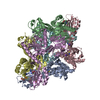


 PDBj
PDBj


 Assembly