Protocol: SINGLE WAVELENGTH / Monochromatic (M) / Laue (L): M / Scattering type: x-ray
Radiation wavelength
Wavelength: 0.9801 Å / Relative weight: 1
Reflection
Resolution: 1.85→65.67 Å / Num. obs: 46925 / % possible obs: 98.8 % / Observed criterion σ(I): 2 / Redundancy: 4.3 % / Rmerge(I) obs: 0.1 / Net I/σ(I): 7.9
Reflection shell
Resolution: 1.85→1.95 Å / Redundancy: 2.9 % / Rmerge(I) obs: 0.53 / Mean I/σ(I) obs: 1.8 / % possible all: 94.5
-
Processing
Software
Name
Version
Classification
REFMAC
5.5.0109
refinement
SCALA
datascaling
Refinement
Method to determine structure: OTHER Starting model: NONE Resolution: 1.85→65.66 Å / Cor.coef. Fo:Fc: 0.944 / Cor.coef. Fo:Fc free: 0.92 / SU B: 4.133 / SU ML: 0.118 / Cross valid method: THROUGHOUT / ESU R: 0.163 / ESU R Free: 0.15 / Stereochemistry target values: MAXIMUM LIKELIHOOD Details: HYDROGENS HAVE BEEN ADDED IN THE RIDING POSITIONS. U VALUES, REFINED INDIVIDUALLY
Rfactor
Num. reflection
% reflection
Selection details
Rfree
0.24269
2365
5.1 %
RANDOM
Rwork
0.19964
-
-
-
obs
0.20178
44413
98.48 %
-
Solvent computation
Ion probe radii: 0.8 Å / Shrinkage radii: 0.8 Å / VDW probe radii: 1.2 Å / Solvent model: MASK
 Movie
Movie Controller

 Yorodumi
 Open data
 Basic information
 Components
 Keywords
 Function and homology information
 Authors
 Citation
 Structure visualization
 Downloads & links
 Other downloads


 PDBj
PDBj
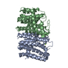
 Assembly

 Mass: 24.305 Da / Num. of mol.: 9 / Source method: obtained synthetically / Formula: Mg
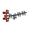
 Mass: 319.229 Da / Num. of mol.: 5 / Source method: obtained synthetically / Formula: C9H23NO7P2 / Comment: medication*YM
 Mass: 18.015 Da / Num. of mol.: 570 / Source method: isolated from a natural source / Formula: H2O
 Sample preparation
 / Beamline: ID14-4 / Type:
  Processing