[English] 日本語
Yorodumi
- PDB-4rh7: Crystal structure of human cytoplasmic dynein 2 motor domain in c... -

+
Open data


ID or keywords:

Loading...

-
Basic information

Entry
Database: PDB / ID: 4rh7
TitleCrystal structure of human cytoplasmic dynein 2 motor domain in complex with ADP.Vi
ComponentsGreen fluorescent protein/Cytoplasmic dynein 2 heavy chain 1
KeywordsMOTOR PROTEIN / AAA+ protein / dynein motor domain
Function / homology
Function and homology information


intraciliary retrograde transport / cilium movement involved in cell motility / 9+2 motile cilium / spinal cord motor neuron differentiation / ciliary tip / Intraflagellar transport / protein localization to cilium / minus-end-directed microtubule motor activity / cytoplasmic dynein complex / non-motile cilium assembly ...intraciliary retrograde transport / cilium movement involved in cell motility / 9+2 motile cilium / spinal cord motor neuron differentiation / ciliary tip / Intraflagellar transport / protein localization to cilium / minus-end-directed microtubule motor activity / cytoplasmic dynein complex / non-motile cilium assembly / coronary vasculature development / dynein light intermediate chain binding / positive regulation of smoothened signaling pathway / dorsal/ventral pattern formation / determination of left/right symmetry / embryonic limb morphogenesis / dynein intermediate chain binding / Golgi organization / axoneme / cytoskeletal motor activity / Hedgehog 'off' state / forebrain development / kidney development / cilium / protein processing / apical part of cell / microtubule / Golgi apparatus / ATP hydrolysis activity / extracellular exosome / ATP binding / plasma membrane
Similarity search - Function
: / Cytoplasmic dynein 2 heavy chain 1, AAA+ ATPase domain / Dynein heavy chain, C-terminal domain / Dynein heavy chain, C-terminal domain, barrel region / Dynein heavy chain C-terminal domain / P-loop containing dynein motor region / Dynein heavy chain, tail / Dynein heavy chain, N-terminal region 1 / Dynein heavy chain / Dynein heavy chain region D6 P-loop domain ...: / Cytoplasmic dynein 2 heavy chain 1, AAA+ ATPase domain / Dynein heavy chain, C-terminal domain / Dynein heavy chain, C-terminal domain, barrel region / Dynein heavy chain C-terminal domain / P-loop containing dynein motor region / Dynein heavy chain, tail / Dynein heavy chain, N-terminal region 1 / Dynein heavy chain / Dynein heavy chain region D6 P-loop domain / Dynein heavy chain, linker / Dynein heavy chain, AAA module D4 / Dynein heavy chain, coiled coil stalk / Dynein heavy chain, hydrolytic ATP-binding dynein motor region / Dynein heavy chain, ATP-binding dynein motor region / Dynein heavy chain AAA lid domain / Dynein heavy chain AAA lid domain superfamily / Dynein heavy chain, domain 2, N-terminal / Dynein heavy chain, linker, subdomain 3 / Dynein heavy chain, AAA1 domain, small subdomain / Dynein heavy chain region D6 P-loop domain / Dynein heavy chain, N-terminal region 2 / Hydrolytic ATP binding site of dynein motor region / Microtubule-binding stalk of dynein motor / P-loop containing dynein motor region D4 / ATP-binding dynein motor region / Dynein heavy chain AAA lid domain / ATPases associated with a variety of cellular activities / AAA+ ATPase domain / P-loop containing nucleoside triphosphate hydrolase
Similarity search - Domain/homology
ADENOSINE-5'-DIPHOSPHATE / ADP ORTHOVANADATE / ADENOSINE-5'-TRIPHOSPHATE / Cytoplasmic dynein 2 heavy chain 1
Similarity search - Component
Biological speciessynthetic construct (others)
Homo sapiens (human)
MethodX-RAY DIFFRACTION / SYNCHROTRON / MIRAS / Resolution: 3.41 Å
AuthorsSchmidt, H. / Zalyte, R. / Urnavicius, L. / Carter, A.P.
CitationJournal: Nature / Year: 2015
Title: Structure of human cytoplasmic dynein-2 primed for its power stroke.
Authors: Schmidt, H. / Zalyte, R. / Urnavicius, L. / Carter, A.P.
History
DepositionOct 1, 2014Deposition site: RCSB / Processing site: RCSB
Revision 1.0Dec 10, 2014Provider: repository / Type: Initial release
Revision 1.1Dec 24, 2014Group: Database references
Revision 1.2Apr 8, 2015Group: Database references
Revision 1.3Aug 2, 2017Group: Refinement description / Source and taxonomy / Category: entity_src_gen / software
Revision 1.4Feb 28, 2024Group: Data collection / Database references / Derived calculations
Category: chem_comp_atom / chem_comp_bond ...chem_comp_atom / chem_comp_bond / database_2 / pdbx_struct_conn_angle / struct_conn / struct_ref_seq_dif / struct_site
Item: _database_2.pdbx_DOI / _database_2.pdbx_database_accession ..._database_2.pdbx_DOI / _database_2.pdbx_database_accession / _pdbx_struct_conn_angle.ptnr1_auth_comp_id / _pdbx_struct_conn_angle.ptnr1_auth_seq_id / _pdbx_struct_conn_angle.ptnr1_label_asym_id / _pdbx_struct_conn_angle.ptnr1_label_atom_id / _pdbx_struct_conn_angle.ptnr1_label_comp_id / _pdbx_struct_conn_angle.ptnr1_label_seq_id / _pdbx_struct_conn_angle.ptnr3_auth_comp_id / _pdbx_struct_conn_angle.ptnr3_auth_seq_id / _pdbx_struct_conn_angle.ptnr3_label_asym_id / _pdbx_struct_conn_angle.ptnr3_label_atom_id / _pdbx_struct_conn_angle.ptnr3_label_comp_id / _pdbx_struct_conn_angle.ptnr3_label_seq_id / _pdbx_struct_conn_angle.value / _struct_conn.pdbx_dist_value / _struct_conn.ptnr1_auth_comp_id / _struct_conn.ptnr1_auth_seq_id / _struct_conn.ptnr1_label_asym_id / _struct_conn.ptnr1_label_atom_id / _struct_conn.ptnr1_label_comp_id / _struct_conn.ptnr1_label_seq_id / _struct_conn.ptnr2_auth_seq_id / _struct_conn.ptnr2_label_asym_id / _struct_ref_seq_dif.details / _struct_site.pdbx_auth_asym_id / _struct_site.pdbx_auth_comp_id / _struct_site.pdbx_auth_seq_id
Remark 650HELIX DETERMINATION METHOD: AUTHOR
Remark 700SHEET DETERMINATION METHOD: AUTHOR

-
Structure visualization

Structure viewerMolecule:
MolmilJmol/JSmol

Downloads & links

-
Assembly

Deposited unit
A: Green fluorescent protein/Cytoplasmic dynein 2 heavy chain 1
hetero molecules


Theoretical massNumber of molelcules
Total (without water)394,9177
Polymers392,9631
Non-polymers1,9546
Water0
1


  • Idetical with deposited unit
  • defined by author
TypeNameSymmetry operationNumber
identity operation1_555x,y,z1
Unit cell
Length a, b, c (Å)136.030, 487.150, 276.460
Angle α, β, γ (deg.)90.00, 90.00, 90.00
Int Tables number20
Space group name H-MC2221

-
Components

#1: Antibody Green fluorescent protein/Cytoplasmic dynein 2 heavy chain 1


Mass: 392963.094 Da / Num. of mol.: 1
Source method: isolated from a genetically manipulated source
Source: (gene. exp.) synthetic construct (others), (gene. exp.) Homo sapiens (human)
Cell line (production host): SF9 / Production host: Spodoptera frugiperda (fall armyworm) / References: UniProt: Q8NCM8
#2: Chemical ChemComp-AOV / ADP ORTHOVANADATE


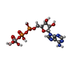
Mass: 544.156 Da / Num. of mol.: 1 / Source method: obtained synthetically / Formula: C10H17N5O14P2V / Comment: energy-carrying molecule analogue*YM
#3: Chemical ChemComp-MG / MAGNESIUM ION


Mass: 24.305 Da / Num. of mol.: 2 / Source method: obtained synthetically / Formula: Mg
#4: Chemical ChemComp-ATP / ADENOSINE-5'-TRIPHOSPHATE / Adenosine triphosphate


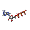
Mass: 507.181 Da / Num. of mol.: 1 / Source method: obtained synthetically / Formula: C10H16N5O13P3 / Comment: ATP, energy-carrying molecule*YM
#5: Chemical ChemComp-ADP / ADENOSINE-5'-DIPHOSPHATE / Adenosine diphosphate


Mass: 427.201 Da / Num. of mol.: 2 / Source method: obtained synthetically / Formula: C10H15N5O10P2 / Comment: ADP, energy-carrying molecule*YM

-
Experimental details

-
Experiment

ExperimentMethod: X-RAY DIFFRACTION / Number of used crystals: 1

-
Sample preparation

CrystalDensity Matthews: 5.83 Å3/Da / Density % sol: 78.89 %
Crystal growTemperature: 292 K / Method: vapor diffusion, hanging drop
Details: mixing equal volumes of protein (8mg/ml) and reservoir solution (4-6 % PEG 6000 and 0.1 M Tris pH 8.0), VAPOR DIFFUSION, HANGING DROP, temperature 292K

-
Data collection

DiffractionMean temperature: 100 K
Diffraction sourceSource: SYNCHROTRON / Site: Diamond / Beamline: I02
DetectorType: PSI PILATUS 6M / Detector: PIXEL / Date: Feb 20, 2014
RadiationProtocol: SINGLE WAVELENGTH / Monochromatic (M) / Laue (L): M / Scattering type: x-ray
Radiation wavelengthRelative weight: 1
ReflectionResolution: 3.4→56.5 Å / Num. obs: 78411 / % possible obs: 62.2 % / Redundancy: 4.1 % / Rsym value: 0.101 / Net I/σ(I): 7.6

-
Processing

Software
NameVersionClassification
SHARPphasing
REFMAC5.8.0073refinement
MOSFLMdata reduction
SCALAdata scaling
RefinementMethod to determine structure: MIRAS / Resolution: 3.41→56.6 Å / Cor.coef. Fo:Fc: 0.924 / Cor.coef. Fo:Fc free: 0.886 / SU B: 35.363 / SU ML: 0.532 / Cross valid method: THROUGHOUT / ESU R Free: 0.643 / Stereochemistry target values: MAXIMUM LIKELIHOOD / Details: HYDROGENS HAVE BEEN ADDED IN THE RIDING POSITIONS
RfactorNum. reflection% reflectionSelection details
Rfree0.28546 3915 5 %RANDOM
Rwork0.23746 ---
obs0.23989 74060 62.16 %-
Solvent computationIon probe radii: 0.8 Å / Shrinkage radii: 0.8 Å / VDW probe radii: 1.2 Å / Solvent model: MASK
Displacement parametersBiso mean: 114.274 Å2
Baniso -1Baniso -2Baniso -3
1-0.05 Å20 Å20 Å2
2---0.28 Å2-0 Å2
3---0.23 Å2
Refinement stepCycle: LAST / Resolution: 3.41→56.6 Å
ProteinNucleic acidLigandSolventTotal
Num. atoms22697 0 119 0 22816
Refine LS restraints
Refine-IDTypeDev idealDev ideal targetNumber
X-RAY DIFFRACTIONr_bond_refined_d0.0120.01923272
X-RAY DIFFRACTIONr_bond_other_d0.0060.0221480
X-RAY DIFFRACTIONr_angle_refined_deg1.5481.96631669
X-RAY DIFFRACTIONr_angle_other_deg0.846349143
X-RAY DIFFRACTIONr_dihedral_angle_1_deg7.26953000
X-RAY DIFFRACTIONr_dihedral_angle_2_deg32.23424.481973
X-RAY DIFFRACTIONr_dihedral_angle_3_deg17.385153715
X-RAY DIFFRACTIONr_dihedral_angle_4_deg15.715121
X-RAY DIFFRACTIONr_chiral_restr0.0730.23633
X-RAY DIFFRACTIONr_gen_planes_refined0.0050.0226721
X-RAY DIFFRACTIONr_gen_planes_other0.0020.025304
X-RAY DIFFRACTIONr_nbd_refined
X-RAY DIFFRACTIONr_nbd_other
X-RAY DIFFRACTIONr_nbtor_refined
X-RAY DIFFRACTIONr_nbtor_other
X-RAY DIFFRACTIONr_xyhbond_nbd_refined
X-RAY DIFFRACTIONr_xyhbond_nbd_other
X-RAY DIFFRACTIONr_metal_ion_refined
X-RAY DIFFRACTIONr_metal_ion_other
X-RAY DIFFRACTIONr_symmetry_vdw_refined
X-RAY DIFFRACTIONr_symmetry_vdw_other
X-RAY DIFFRACTIONr_symmetry_hbond_refined
X-RAY DIFFRACTIONr_symmetry_hbond_other
X-RAY DIFFRACTIONr_symmetry_metal_ion_refined
X-RAY DIFFRACTIONr_symmetry_metal_ion_other
X-RAY DIFFRACTIONr_mcbond_it8.24112.30712015
X-RAY DIFFRACTIONr_mcbond_other8.24112.30612014
X-RAY DIFFRACTIONr_mcangle_it13.70618.45115010
X-RAY DIFFRACTIONr_mcangle_other13.67718.43915011
X-RAY DIFFRACTIONr_scbond_it8.50811.82811257
X-RAY DIFFRACTIONr_scbond_other8.49311.81211256
X-RAY DIFFRACTIONr_scangle_it
X-RAY DIFFRACTIONr_scangle_other14.65617.58516656
X-RAY DIFFRACTIONr_long_range_B_refined25.39101750
X-RAY DIFFRACTIONr_long_range_B_other25.39101751
X-RAY DIFFRACTIONr_rigid_bond_restr
X-RAY DIFFRACTIONr_sphericity_free
X-RAY DIFFRACTIONr_sphericity_bonded
LS refinement shellResolution: 3.406→3.494 Å / Total num. of bins used: 20
RfactorNum. reflection% reflection
Rfree0.541 14 -
Rwork0.345 209 -
obs--2.44 %

+
About Yorodumi

-
News

-
Feb 9, 2022. New format data for meta-information of EMDB entries

New format data for meta-information of EMDB entries

  • Version 3 of the EMDB header file is now the official format.
  • The previous official version 1.9 will be removed from the archive.

Related info.:EMDB header

External links:wwPDB to switch to version 3 of the EMDB data model

-
Aug 12, 2020. Covid-19 info

Covid-19 info

URL: https://pdbj.org/emnavi/covid19.php

New page: Covid-19 featured information page in EM Navigator.

Related info.:Covid-19 info / Mar 5, 2020. Novel coronavirus structure data

+
Mar 5, 2020. Novel coronavirus structure data

Novel coronavirus structure data

Related info.:Yorodumi Speices / Aug 12, 2020. Covid-19 info

External links:COVID-19 featured content - PDBj / Molecule of the Month (242):Coronavirus Proteases

+
Jan 31, 2019. EMDB accession codes are about to change! (news from PDBe EMDB page)

EMDB accession codes are about to change! (news from PDBe EMDB page)

  • The allocation of 4 digits for EMDB accession codes will soon come to an end. Whilst these codes will remain in use, new EMDB accession codes will include an additional digit and will expand incrementally as the available range of codes is exhausted. The current 4-digit format prefixed with “EMD-” (i.e. EMD-XXXX) will advance to a 5-digit format (i.e. EMD-XXXXX), and so on. It is currently estimated that the 4-digit codes will be depleted around Spring 2019, at which point the 5-digit format will come into force.
  • The EM Navigator/Yorodumi systems omit the EMD- prefix.

Related info.:Q: What is EMD? / ID/Accession-code notation in Yorodumi/EM Navigator

External links:EMDB Accession Codes are Changing Soon! / Contact to PDBj

+
Jul 12, 2017. Major update of PDB

Major update of PDB

  • wwPDB released updated PDB data conforming to the new PDBx/mmCIF dictionary.
  • This is a major update changing the version number from 4 to 5, and with Remediation, in which all the entries are updated.
  • In this update, many items about electron microscopy experimental information are reorganized (e.g. em_software).
  • Now, EM Navigator and Yorodumi are based on the updated data.

External links:wwPDB Remediation / Enriched Model Files Conforming to OneDep Data Standards Now Available in the PDB FTP Archive

-
Yorodumi

Thousand views of thousand structures

  • Yorodumi is a browser for structure data from EMDB, PDB, SASBDB, etc.
  • This page is also the successor to EM Navigator detail page, and also detail information page/front-end page for Omokage search.
  • The word "yorodu" (or yorozu) is an old Japanese word meaning "ten thousand". "mi" (miru) is to see.

Related info.:EMDB / PDB / SASBDB / Comparison of 3 databanks / Yorodumi Search / Aug 31, 2016. New EM Navigator & Yorodumi / Yorodumi Papers / Jmol/JSmol / Function and homology information / Changes in new EM Navigator and Yorodumi

Read more