 Movie
Movie Controller


+ Open data

- Basic information
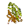
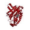
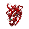
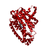
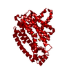
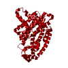
| Entry | Database: PDB / ID: 4qxs | ||||||
|---|---|---|---|---|---|---|---|
| Title | Crystal structure of human FPPS in complex with WC01088 | ||||||
 Components Components | Farnesyl pyrophosphate synthase Dimethylallyltranstransferase Dimethylallyltranstransferase | ||||||
 Keywords Keywords | TRANSFERASE/TRANSFERASE INHIBITOR / TRANSFERASE-TRANSFERASE INHIBITOR complex | ||||||
| Function / homology |  Function and homology information Function and homology informationgeranyl diphosphate biosynthetic process / dimethylallyltranstransferase / (2E,6E)-farnesyl diphosphate synthase / Cholesterol biosynthesis / farnesyl diphosphate biosynthetic process / dimethylallyltranstransferase activity / geranyltranstransferase activity / cholesterol biosynthetic process / Activation of gene expression by SREBF (SREBP) / RNA binding ...geranyl diphosphate biosynthetic process / dimethylallyltranstransferase / (2E,6E)-farnesyl diphosphate synthase / Cholesterol biosynthesis / farnesyl diphosphate biosynthetic process / dimethylallyltranstransferase activity / geranyltranstransferase activity / cholesterol biosynthetic process / Activation of gene expression by SREBF (SREBP) / RNA binding / nucleoplasm / metal ion binding / cytosol / cytoplasmSimilarity search - Function | ||||||
| Biological species |  Homo sapiens (human) Homo sapiens (human) | ||||||
| Method | X-RAY DIFFRACTION / SYNCHROTRON / FOURIER SYNTHESIS / Resolution: 1.9 Å | ||||||
 Authors Authors | Park, J. / Zielinski, M. / Weiling, C. / Tsantrizos, Y.S. / Berghuis, A.M. | ||||||
 Citation Citation | Journal: Bioorg.Med.Chem.Lett. / Year: 2015 Title: Probing the molecular and structural elements of ligands binding to the active site versus an allosteric pocket of the human farnesyl pyrophosphate synthase. Authors: Gritzalis, D. / Park, J. / Chiu, W. / Cho, H. / Lin, Y.S. / De Schutter, J.W. / Lacbay, C.M. / Zielinski, M. / Berghuis, A.M. / Tsantrizos, Y.S. | ||||||
| History |
|
- Structure visualization
| Structure viewer | Molecule: MolmilJmol/JSmol |
|---|
- Downloads & links
-Download
| PDBx/mmCIF format | 4qxs.cif.gz | 156.9 KB | Display | PDBx/mmCIF format |
|---|---|---|---|---|
| PDB format | pdb4qxs.ent.gz | 122.8 KB | Display | PDB format |
| PDBx/mmJSON format | 4qxs.json.gz | Tree view | PDBx/mmJSON format | |
| Others |  Other downloads Other downloads |
-Validation report
| Arichive directory | https://data.pdbj.org/pub/pdb/validation_reports/qx/4qxsftp://data.pdbj.org/pub/pdb/validation_reports/qx/4qxs | HTTPS FTP |
|---|
-Related structure data
| Related structure data |  2f7mS S: Starting model for refinement |
|---|---|
| Similar structure data |


-Links
 PDBj
PDBj

- Assembly
| Deposited unit | 
| |||||||||
|---|---|---|---|---|---|---|---|---|---|---|
| 1 | 
| |||||||||
| Unit cell |
| |||||||||
| Components on special symmetry positions |
|
-Components
| #1: Protein | Dimethylallyltranstransferase / FPP synthase / FPS / (2E / 6E)-farnesyl diphosphate synthase / Dimethylallyltranstransferase / ...FPP synthase / FPS / (2E / 6E)-farnesyl diphosphate synthase / Dimethylallyltranstransferase / Farnesyl diphosphate synthase / Geranyltranstransferase Mass: 43144.980 Da / Num. of mol.: 1 / Fragment: UNP residues 67-419 Source method: isolated from a genetically manipulated source Source: (gene. exp.) Homo sapiens (human) / Gene: FDPS, FPS, KIAA1293 / Production host:  Escherichia coli (E. coli) Escherichia coli (E. coli)References: UniProt: P14324, (2E,6E)-farnesyl diphosphate synthase, dimethylallyltranstransferase | ||||||
|---|---|---|---|---|---|---|---|
| #2: Chemical | Phosphate  Mass: 94.971 Da / Num. of mol.: 2 / Source method: obtained synthetically / Formula: PO4 Mass: 94.971 Da / Num. of mol.: 2 / Source method: obtained synthetically / Formula: PO4#3: Chemical | ChemComp-WC1 / ( |   Mass: 451.390 Da / Num. of mol.: 1 / Source method: obtained synthetically / Formula: C21H27NO6P2 Mass: 451.390 Da / Num. of mol.: 1 / Source method: obtained synthetically / Formula: C21H27NO6P2#4: Chemical | ChemComp-GOL / | Glycerol  Mass: 92.094 Da / Num. of mol.: 1 / Source method: obtained synthetically / Formula: C3H8O3 Mass: 92.094 Da / Num. of mol.: 1 / Source method: obtained synthetically / Formula: C3H8O3#5: Water | ChemComp-HOH / | Water Mass: 18.015 Da / Num. of mol.: 144 / Source method: isolated from a natural source / Formula: H2O Mass: 18.015 Da / Num. of mol.: 144 / Source method: isolated from a natural source / Formula: H2O |
-Experimental details
-Experiment
| Experiment | Method: X-RAY DIFFRACTION / Number of used crystals: 1 |
|---|
- Sample preparation
| Crystal | Density Matthews: 2.76 Å3/Da / Density % sol: 55.5 % |
|---|---|
| Crystal grow | Temperature: 295 K / Method: vapor diffusion, sitting drop / pH: 8.5 Details: 1.6 M ammonium phosphate, 20% glycerol, 0.08 M Tris-HCl, pH 8.5, VAPOR DIFFUSION, SITTING DROP, temperature 295K |
-Data collection
| Diffraction | Mean temperature: 100 K |
|---|---|
| Diffraction source | Source: SYNCHROTRON / Site: CLSI  / Beamline: 08ID-1 / Wavelength: 0.97949 Å / Beamline: 08ID-1 / Wavelength: 0.97949 Å |
| Detector | Type: RAYONIX MX-300 / Detector: CCD / Date: Jul 8, 2014 |
| Radiation | Monochromator: ACCEL/Bruker double crystal Si(111) / Protocol: SINGLE WAVELENGTH / Monochromatic (M) / Laue (L): M / Scattering type: x-ray |
| Radiation wavelength | Wavelength: 0.97949 Å / Relative weight: 1 |
| Reflection | Resolution: 1.9→55.21 Å / Num. all: 38434 / Num. obs: 38434 / % possible obs: 99.4 % / Redundancy: 9.7 % / Biso Wilson estimate: 36.5 Å2 / Rmerge(I) obs: 0.041 / Net I/σ(I): 28.2 |
| Reflection shell | Resolution: 1.9→1.95 Å / Redundancy: 9.6 % / Rmerge(I) obs: 1.177 / Mean I/σ(I) obs: 2.9 / Num. unique all: 2768 / % possible all: 98.6 |
- Processing
| Software |
| |||||||||||||||||||||||||||||||||||||||||||||||||||||||||||||||||||||||||||||||||||||||||||||||||||||||||||||||||||||||||||||||||||||||||||||||||||||||||||||||||||||||||||||||
|---|---|---|---|---|---|---|---|---|---|---|---|---|---|---|---|---|---|---|---|---|---|---|---|---|---|---|---|---|---|---|---|---|---|---|---|---|---|---|---|---|---|---|---|---|---|---|---|---|---|---|---|---|---|---|---|---|---|---|---|---|---|---|---|---|---|---|---|---|---|---|---|---|---|---|---|---|---|---|---|---|---|---|---|---|---|---|---|---|---|---|---|---|---|---|---|---|---|---|---|---|---|---|---|---|---|---|---|---|---|---|---|---|---|---|---|---|---|---|---|---|---|---|---|---|---|---|---|---|---|---|---|---|---|---|---|---|---|---|---|---|---|---|---|---|---|---|---|---|---|---|---|---|---|---|---|---|---|---|---|---|---|---|---|---|---|---|---|---|---|---|---|---|---|---|---|---|
| Refinement | Method to determine structure: FOURIER SYNTHESIS Starting model: PDB ENTRY 2F7M Resolution: 1.9→55.21 Å / Cor.coef. Fo:Fc: 0.972 / Cor.coef. Fo:Fc free: 0.955 / SU B: 7.827 / SU ML: 0.106 / Cross valid method: THROUGHOUT / ESU R: 0.112 / ESU R Free: 0.112 / Stereochemistry target values: MAXIMUM LIKELIHOOD / Details: HYDROGENS HAVE BEEN ADDED IN THE RIDING POSITIONS
| |||||||||||||||||||||||||||||||||||||||||||||||||||||||||||||||||||||||||||||||||||||||||||||||||||||||||||||||||||||||||||||||||||||||||||||||||||||||||||||||||||||||||||||||
| Solvent computation | Ion probe radii: 0.8 Å / Shrinkage radii: 0.8 Å / VDW probe radii: 1.2 Å / Solvent model: MASK | |||||||||||||||||||||||||||||||||||||||||||||||||||||||||||||||||||||||||||||||||||||||||||||||||||||||||||||||||||||||||||||||||||||||||||||||||||||||||||||||||||||||||||||||
| Displacement parameters | Biso mean: 59.447 Å2
| |||||||||||||||||||||||||||||||||||||||||||||||||||||||||||||||||||||||||||||||||||||||||||||||||||||||||||||||||||||||||||||||||||||||||||||||||||||||||||||||||||||||||||||||
| Refinement step | Cycle: LAST / Resolution: 1.9→55.21 Å
| |||||||||||||||||||||||||||||||||||||||||||||||||||||||||||||||||||||||||||||||||||||||||||||||||||||||||||||||||||||||||||||||||||||||||||||||||||||||||||||||||||||||||||||||
| Refine LS restraints |
| |||||||||||||||||||||||||||||||||||||||||||||||||||||||||||||||||||||||||||||||||||||||||||||||||||||||||||||||||||||||||||||||||||||||||||||||||||||||||||||||||||||||||||||||
| LS refinement shell | Resolution: 1.9→1.949 Å / Total num. of bins used: 20
| |||||||||||||||||||||||||||||||||||||||||||||||||||||||||||||||||||||||||||||||||||||||||||||||||||||||||||||||||||||||||||||||||||||||||||||||||||||||||||||||||||||||||||||||
| Refinement TLS params. | Method: refined / Refine-ID: X-RAY DIFFRACTION
| |||||||||||||||||||||||||||||||||||||||||||||||||||||||||||||||||||||||||||||||||||||||||||||||||||||||||||||||||||||||||||||||||||||||||||||||||||||||||||||||||||||||||||||||
| Refinement TLS group |
|
