 Movie
Movie Controller


+ Open data

- Basic information
| Entry | Database: PDB / ID: 4qh7 | ||||||
|---|---|---|---|---|---|---|---|
| Title | LC8 - Ana2 (159-168) Complex | ||||||
 Components Components |
| ||||||
 Keywords Keywords |  MOTOR PROTEIN / LC8 fold Dimer / Target dimerization / Ana2 / Cellular MOTOR PROTEIN / LC8 fold Dimer / Target dimerization / Ana2 / Cellular | ||||||
| Function / homology |  Function and homology information Function and homology informationciliary basal body organization / spermatid nucleus elongation / chaeta morphogenesis / positive regulation of neuron remodeling / Macroautophagy / Aggrephagy / wing disc development / HSP90 chaperone cycle for steroid hormone receptors (SHR) in the presence of ligand / COPI-mediated anterograde transport / COPI-independent Golgi-to-ER retrograde traffic ...ciliary basal body organization / spermatid nucleus elongation / chaeta morphogenesis / positive regulation of neuron remodeling / Macroautophagy / Aggrephagy / wing disc development / HSP90 chaperone cycle for steroid hormone receptors (SHR) in the presence of ligand / COPI-mediated anterograde transport / COPI-independent Golgi-to-ER retrograde traffic / centriole assembly / centriole-centriole cohesion / : / chaeta development / sperm individualization / microtubule anchoring at centrosome / imaginal disc-derived wing morphogenesis / asymmetric neuroblast division / Neutrophil degranulation / dynein complex / cytoplasmic dynein complex / dynein light intermediate chain binding / dynein intermediate chain binding / oogenesis / establishment of mitotic spindle orientation / centriole replication / actin filament bundle assembly / centriole / ciliary basal body / autophagy / disordered domain specific binding / spermatogenesis / microtubule / centrosome / protein homodimerization activity / protein-containing complex / cytoplasmSimilarity search - Function | ||||||
| Biological species |  Drosophila melanogaster (fruit fly) Drosophila melanogaster (fruit fly) | ||||||
| Method | X-RAY DIFFRACTION / SYNCHROTRON / MOLECULAR REPLACEMENT / Resolution: 1.829 Å | ||||||
 Authors Authors | Slevin, L.K. / Romes, E.R. / Slep, K.C. | ||||||
 Citation Citation | Journal: J.Biol.Chem. / Year: 2014 Title: The Mechanism of Dynein Light Chain LC8-mediated Oligomerization of the Ana2 Centriole Duplication Factor. Authors: Slevin, L.K. / Romes, E.M. / Dandulakis, M.G. / Slep, K.C. | ||||||
| History |
|
- Structure visualization
| Structure viewer | Molecule: MolmilJmol/JSmol |
|---|
- Downloads & links
-Download
| PDBx/mmCIF format | 4qh7.cif.gz | 95.2 KB | Display | PDBx/mmCIF format |
|---|---|---|---|---|
| PDB format | pdb4qh7.ent.gz | 73.2 KB | Display | PDB format |
| PDBx/mmJSON format | 4qh7.json.gz | Tree view | PDBx/mmJSON format | |
| Others |  Other downloads Other downloads |
-Validation report
| Arichive directory | https://data.pdbj.org/pub/pdb/validation_reports/qh/4qh7ftp://data.pdbj.org/pub/pdb/validation_reports/qh/4qh7 | HTTPS FTP |
|---|
-Related structure data
| Related structure data |  4qh8C 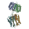 2pg1S C: citing same article ( S: Starting model for refinement |
|---|---|
| Similar structure data |


-Links
 PDBj
PDBj

- Assembly
| Deposited unit | 
| ||||||||
|---|---|---|---|---|---|---|---|---|---|
| 1 | 
| ||||||||
| 2 | 
| ||||||||
| Unit cell |
|
-Components
| #1: Protein | Mass: 10800.304 Da / Num. of mol.: 4 / Fragment: LC8 Source method: isolated from a genetically manipulated source Source: (gene. exp.) Drosophila melanogaster (fruit fly) / Gene: ctp, Cdlc1, ddlc1, CG6998 / Plasmid: pGEX-6P-2 / Production host:  Escherichia coli (E. coli) / Strain (production host): BL21 DE3 / References: UniProt: Q24117 Escherichia coli (E. coli) / Strain (production host): BL21 DE3 / References: UniProt: Q24117#2: Protein/peptide | Mass: 1283.365 Da / Num. of mol.: 4 / Fragment: Ana2 T159-P168 / Source method: obtained synthetically Details: Sequence T159-P168 occurs naturally in Drosophila melanogaster. Peptide synthesized is NYTICAGTQTDP with C-terminal amide. Source: (synth.) Drosophila melanogaster (fruit fly) / References: UniProt: Q9XZ31#3: Water | ChemComp-HOH / | Water Mass: 18.015 Da / Num. of mol.: 310 / Source method: isolated from a natural source / Formula: H2O Mass: 18.015 Da / Num. of mol.: 310 / Source method: isolated from a natural source / Formula: H2O |
|---|
-Experimental details
-Experiment
| Experiment | Method: X-RAY DIFFRACTION / Number of used crystals: 1 |
|---|
- Sample preparation
| Crystal | Density Matthews: 2.26 Å3/Da / Density % sol: 45.52 % |
|---|---|
| Crystal grow | Temperature: 293.15 K / Method: vapor diffusion, hanging drop / pH: 6.5 Details: 0.3M magnesium acetate, 0.1M sodium cacodylate, 26% PEG 8000, pH 6.5, VAPOR DIFFUSION, HANGING DROP, temperature 293.15K |
-Data collection
| Diffraction | Mean temperature: 100 K |
|---|---|
| Diffraction source | Source: SYNCHROTRON / Site: APS  / Beamline: 22-ID / Wavelength: 1 Å / Beamline: 22-ID / Wavelength: 1 Å |
| Detector | Type: MARMOSAIC 300 mm CCD / Detector: CCD / Date: Sep 14, 2012 |
| Radiation | Monochromator: Double crystal - liquid nitrogen-cooled / Protocol: SINGLE WAVELENGTH / Monochromatic (M) / Laue (L): M / Scattering type: x-ray |
| Radiation wavelength | Wavelength: 1 Å / Relative weight: 1 |
| Reflection | Resolution: 1.83→50 Å / Num. all: 108273 / Num. obs: 37488 / % possible obs: 95.1 % / Observed criterion σ(F): 2 / Observed criterion σ(I): 2 / Rsym value: 0.08 / Net I/σ(I): 13.7 |
| Reflection shell | Resolution: 1.83→1.9 Å / Mean I/σ(I) obs: 2.4 / Rsym value: 0.37 / % possible all: 95.2 |
- Processing
| Software |
| |||||||||||||||||||||||||||||||||||||||||||||||||||||||||||||||||||||||||||||||||||||||||||||||||||||||||
|---|---|---|---|---|---|---|---|---|---|---|---|---|---|---|---|---|---|---|---|---|---|---|---|---|---|---|---|---|---|---|---|---|---|---|---|---|---|---|---|---|---|---|---|---|---|---|---|---|---|---|---|---|---|---|---|---|---|---|---|---|---|---|---|---|---|---|---|---|---|---|---|---|---|---|---|---|---|---|---|---|---|---|---|---|---|---|---|---|---|---|---|---|---|---|---|---|---|---|---|---|---|---|---|---|---|---|
| Refinement | Method to determine structure: MOLECULAR REPLACEMENT Starting model: 2PG1 Resolution: 1.829→44.629 Å / SU ML: 0.15 / σ(F): 0 / Phase error: 19.33 / Stereochemistry target values: ML
| |||||||||||||||||||||||||||||||||||||||||||||||||||||||||||||||||||||||||||||||||||||||||||||||||||||||||
| Solvent computation | Shrinkage radii: 0.9 Å / VDW probe radii: 1.11 Å / Solvent model: FLAT BULK SOLVENT MODEL | |||||||||||||||||||||||||||||||||||||||||||||||||||||||||||||||||||||||||||||||||||||||||||||||||||||||||
| Refinement step | Cycle: LAST / Resolution: 1.829→44.629 Å
| |||||||||||||||||||||||||||||||||||||||||||||||||||||||||||||||||||||||||||||||||||||||||||||||||||||||||
| Refine LS restraints |
| |||||||||||||||||||||||||||||||||||||||||||||||||||||||||||||||||||||||||||||||||||||||||||||||||||||||||
| LS refinement shell |
|
