 Movie
Movie Controller

[English] 日本語
 Yorodumi
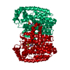
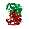
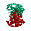
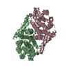
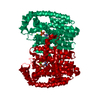
- PDB-4nfj: Crystal structure of human FPPS in complex with magnesium, JDS051... -
+ Open data

- Basic information
| Entry | Database: PDB / ID: 4nfj | ||||||
|---|---|---|---|---|---|---|---|
| Title | Crystal structure of human FPPS in complex with magnesium, JDS05120, and sulfate | ||||||
 Components Components | Farnesyl pyrophosphate synthase Dimethylallyltranstransferase Dimethylallyltranstransferase | ||||||
 Keywords Keywords | Transferase/Transferase inhibitor / Transferase-Transferase inhibitor complex | ||||||
| Function / homology |  Function and homology information Function and homology informationgeranyl diphosphate biosynthetic process / dimethylallyltranstransferase / (2E,6E)-farnesyl diphosphate synthase / Cholesterol biosynthesis / farnesyl diphosphate biosynthetic process / dimethylallyltranstransferase activity / geranyltranstransferase activity / cholesterol biosynthetic process / Activation of gene expression by SREBF (SREBP) / RNA binding ...geranyl diphosphate biosynthetic process / dimethylallyltranstransferase / (2E,6E)-farnesyl diphosphate synthase / Cholesterol biosynthesis / farnesyl diphosphate biosynthetic process / dimethylallyltranstransferase activity / geranyltranstransferase activity / cholesterol biosynthetic process / Activation of gene expression by SREBF (SREBP) / RNA binding / nucleoplasm / metal ion binding / cytosol / cytoplasmSimilarity search - Function | ||||||
| Biological species |  Homo sapiens (human) Homo sapiens (human) | ||||||
| Method | X-RAY DIFFRACTION / SYNCHROTRON / FOURIER SYNTHESIS / Resolution: 2.05 Å | ||||||
 Authors Authors | Park, J. / De Schutter, J.W. / Tsantrizos, Y.S. / Berghuis, A.M. | ||||||
 Citation Citation | Journal: PLoS ONE / Year: 2017 Title: Crystallographic and thermodynamic characterization of phenylaminopyridine bisphosphonates binding to human farnesyl pyrophosphate synthase. Authors: Park, J. / Rodionov, D. / De Schutter, J.W. / Lin, Y.S. / Tsantrizos, Y.S. / Berghuis, A.M. | ||||||
| History |
|
- Structure visualization
| Structure viewer | Molecule: MolmilJmol/JSmol |
|---|
- Downloads & links
-Download
| PDBx/mmCIF format | 4nfj.cif.gz | 155.7 KB | Display | PDBx/mmCIF format |
|---|---|---|---|---|
| PDB format | pdb4nfj.ent.gz | 120.9 KB | Display | PDB format |
| PDBx/mmJSON format | 4nfj.json.gz | Tree view | PDBx/mmJSON format | |
| Others |  Other downloads Other downloads |
-Validation report
| Arichive directory | https://data.pdbj.org/pub/pdb/validation_reports/nf/4nfjftp://data.pdbj.org/pub/pdb/validation_reports/nf/4nfj | HTTPS FTP |
|---|
-Related structure data
| Related structure data |  4nfiC  4nfkC  4pvxC  4pvyC  4demS C: citing same article ( S: Starting model for refinement |
|---|---|
| Similar structure data |


-Links
 PDBj
PDBj

- Assembly
| Deposited unit | 
| ||||||||
|---|---|---|---|---|---|---|---|---|---|
| 1 | 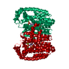
| ||||||||
| Unit cell |
|
-Components
| #1: Protein | Dimethylallyltranstransferase / FPP synthase / FPS / (2E / 6E)-farnesyl diphosphate synthase / Dimethylallyltranstransferase / ...FPP synthase / FPS / (2E / 6E)-farnesyl diphosphate synthase / Dimethylallyltranstransferase / Farnesyl diphosphate synthase / Geranyltranstransferase Mass: 43144.980 Da / Num. of mol.: 1 Source method: isolated from a genetically manipulated source Source: (gene. exp.) Homo sapiens (human) / Gene: FDPS, FPS, KIAA1293 / Production host:  Escherichia coli (E. coli) Escherichia coli (E. coli)References: UniProt: P14324, (2E,6E)-farnesyl diphosphate synthase, dimethylallyltranstransferase | ||||
|---|---|---|---|---|---|
| #2: Chemical | ChemComp-JD5 / [({  Mass: 400.260 Da / Num. of mol.: 1 / Source method: obtained synthetically / Formula: C15H18N2O7P2 Mass: 400.260 Da / Num. of mol.: 1 / Source method: obtained synthetically / Formula: C15H18N2O7P2 | ||||
| #3: Chemical |   Mass: 24.305 Da / Num. of mol.: 3 / Source method: obtained synthetically / Formula: Mg Mass: 24.305 Da / Num. of mol.: 3 / Source method: obtained synthetically / Formula: Mg#4: Chemical | ChemComp-SO4 / | Sulfate  Mass: 96.063 Da / Num. of mol.: 1 / Source method: obtained synthetically / Formula: SO4 Mass: 96.063 Da / Num. of mol.: 1 / Source method: obtained synthetically / Formula: SO4#5: Water | ChemComp-HOH / | Water Mass: 18.015 Da / Num. of mol.: 105 / Source method: isolated from a natural source / Formula: H2O Mass: 18.015 Da / Num. of mol.: 105 / Source method: isolated from a natural source / Formula: H2O |
-Experimental details
-Experiment
| Experiment | Method: X-RAY DIFFRACTION / Number of used crystals: 1 |
|---|
- Sample preparation
| Crystal | Density Matthews: 2.43 Å3/Da / Density % sol: 49.48 % |
|---|---|
| Crystal grow | Temperature: 295 K / Method: vapor diffusion, sitting drop / pH: 4.6 Details: 25.5% PEG MME 2000, 0.17M ammonium sulfate, 15% glycerol, 0.09M sodium acetate, pH 4.6, VAPOR DIFFUSION, SITTING DROP, temperature 295K |
-Data collection
| Diffraction | Mean temperature: 100 K |
|---|---|
| Diffraction source | Source: SYNCHROTRON / Site: CLSI  / Beamline: 08ID-1 / Wavelength: 0.97949 Å / Beamline: 08ID-1 / Wavelength: 0.97949 Å |
| Detector | Type: RAYONIX MX-300 / Detector: CCD / Date: Feb 20, 2013 |
| Radiation | Monochromator: ACCEL/BRUKER DCM / Protocol: SINGLE WAVELENGTH / Monochromatic (M) / Laue (L): M / Scattering type: x-ray |
| Radiation wavelength | Wavelength: 0.97949 Å / Relative weight: 1 |
| Reflection | Resolution: 2.05→50 Å / Num. all: 27177 / Num. obs: 27177 / % possible obs: 99.4 % / Redundancy: 9.5 % / Rmerge(I) obs: 0.06 / Net I/σ(I): 31.3 |
| Reflection shell | Resolution: 2.05→2.09 Å / Redundancy: 9.3 % / Rmerge(I) obs: 0.472 / Mean I/σ(I) obs: 4 / Num. unique all: 1311 / % possible all: 98.6 |
- Processing
| Software |
| |||||||||||||||||||||||||||||||||||||||||||||||||||||||||||||||||||||||||||||||||||||||||||||||||||||||||||||||||||||||||||||
|---|---|---|---|---|---|---|---|---|---|---|---|---|---|---|---|---|---|---|---|---|---|---|---|---|---|---|---|---|---|---|---|---|---|---|---|---|---|---|---|---|---|---|---|---|---|---|---|---|---|---|---|---|---|---|---|---|---|---|---|---|---|---|---|---|---|---|---|---|---|---|---|---|---|---|---|---|---|---|---|---|---|---|---|---|---|---|---|---|---|---|---|---|---|---|---|---|---|---|---|---|---|---|---|---|---|---|---|---|---|---|---|---|---|---|---|---|---|---|---|---|---|---|---|---|---|---|
| Refinement | Method to determine structure: FOURIER SYNTHESIS Starting model: PDB ENTRY 4DEM Resolution: 2.05→50 Å / Cor.coef. Fo:Fc: 0.97 / Cor.coef. Fo:Fc free: 0.956 / SU B: 10.68 / SU ML: 0.135 / Cross valid method: THROUGHOUT / ESU R: 0.161 / ESU R Free: 0.148 / Stereochemistry target values: MAXIMUM LIKELIHOOD / Details: HYDROGENS HAVE BEEN ADDED IN THE RIDING POSITIONS
| |||||||||||||||||||||||||||||||||||||||||||||||||||||||||||||||||||||||||||||||||||||||||||||||||||||||||||||||||||||||||||||
| Solvent computation | Ion probe radii: 0.8 Å / Shrinkage radii: 0.8 Å / VDW probe radii: 1.2 Å / Solvent model: MASK | |||||||||||||||||||||||||||||||||||||||||||||||||||||||||||||||||||||||||||||||||||||||||||||||||||||||||||||||||||||||||||||
| Displacement parameters | Biso mean: 46.148 Å2
| |||||||||||||||||||||||||||||||||||||||||||||||||||||||||||||||||||||||||||||||||||||||||||||||||||||||||||||||||||||||||||||
| Refinement step | Cycle: LAST / Resolution: 2.05→50 Å
| |||||||||||||||||||||||||||||||||||||||||||||||||||||||||||||||||||||||||||||||||||||||||||||||||||||||||||||||||||||||||||||
| Refine LS restraints |
| |||||||||||||||||||||||||||||||||||||||||||||||||||||||||||||||||||||||||||||||||||||||||||||||||||||||||||||||||||||||||||||
| LS refinement shell | Resolution: 2.051→2.104 Å / Total num. of bins used: 20
| |||||||||||||||||||||||||||||||||||||||||||||||||||||||||||||||||||||||||||||||||||||||||||||||||||||||||||||||||||||||||||||
| Refinement TLS params. | Method: refined / Refine-ID: X-RAY DIFFRACTION
| |||||||||||||||||||||||||||||||||||||||||||||||||||||||||||||||||||||||||||||||||||||||||||||||||||||||||||||||||||||||||||||
| Refinement TLS group |
|
