+ Open data
- Basic information
| Entry | Database: PDB / ID: 4mth | ||||||
|---|---|---|---|---|---|---|---|
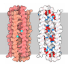
| Title | Crystal structure of mature human RegIIIalpha | ||||||
 Components Components | Regenerating islet-derived protein 3-alpha 15 kDa form | ||||||
 Keywords Keywords |  ANTIMICROBIAL PROTEIN / HIP/PAP / REGIII-GAMMA / ANTIMICROBIAL PROTEIN / HIP/PAP / REGIII-GAMMA /  C-TYPE LECTIN C-TYPE LECTIN | ||||||
| Function / homology |  Function and homology information Function and homology informationdisruption of cell wall in another organism / positive regulation of detection of glucose / response to symbiotic bacterium / positive regulation of keratinocyte proliferation / negative regulation of keratinocyte differentiation / negative regulation of inflammatory response to wounding /  oligosaccharide binding / oligosaccharide binding /  peptidoglycan binding / heterophilic cell-cell adhesion via plasma membrane cell adhesion molecules / peptidoglycan binding / heterophilic cell-cell adhesion via plasma membrane cell adhesion molecules /  Antimicrobial peptides ...disruption of cell wall in another organism / positive regulation of detection of glucose / response to symbiotic bacterium / positive regulation of keratinocyte proliferation / negative regulation of keratinocyte differentiation / negative regulation of inflammatory response to wounding / Antimicrobial peptides ...disruption of cell wall in another organism / positive regulation of detection of glucose / response to symbiotic bacterium / positive regulation of keratinocyte proliferation / negative regulation of keratinocyte differentiation / negative regulation of inflammatory response to wounding /  oligosaccharide binding / oligosaccharide binding /  peptidoglycan binding / heterophilic cell-cell adhesion via plasma membrane cell adhesion molecules / peptidoglycan binding / heterophilic cell-cell adhesion via plasma membrane cell adhesion molecules /  Antimicrobial peptides / positive regulation of wound healing / acute-phase response / Antimicrobial peptides / positive regulation of wound healing / acute-phase response /  hormone activity / response to peptide hormone / response to wounding / negative regulation of inflammatory response / antimicrobial humoral immune response mediated by antimicrobial peptide / hormone activity / response to peptide hormone / response to wounding / negative regulation of inflammatory response / antimicrobial humoral immune response mediated by antimicrobial peptide /  signaling receptor activity / signaling receptor activity /  carbohydrate binding / positive regulation of cell population proliferation / carbohydrate binding / positive regulation of cell population proliferation /  extracellular space / extracellular region / identical protein binding / extracellular space / extracellular region / identical protein binding /  cytoplasm cytoplasmSimilarity search - Function | ||||||
| Biological species |   Homo sapiens (human) Homo sapiens (human) | ||||||
| Method |  X-RAY DIFFRACTION / X-RAY DIFFRACTION /  SYNCHROTRON / SYNCHROTRON /  MOLECULAR REPLACEMENT / MOLECULAR REPLACEMENT /  molecular replacement / Resolution: 1.47 Å molecular replacement / Resolution: 1.47 Å | ||||||
 Authors Authors | Derebe, M.G. | ||||||
 Citation Citation |  Journal: Nature / Year: 2014 Journal: Nature / Year: 2014Title: Antibacterial membrane attack by a pore-forming intestinal C-type lectin. Authors: Sohini Mukherjee / Hui Zheng / Mehabaw G Derebe / Keith M Callenberg / Carrie L Partch / Darcy Rollins / Daniel C Propheter / Josep Rizo / Michael Grabe / Qiu-Xing Jiang / Lora V Hooper /  Abstract: Human body-surface epithelia coexist in close association with complex bacterial communities and are protected by a variety of antibacterial proteins. C-type lectins of the RegIII family are ...Human body-surface epithelia coexist in close association with complex bacterial communities and are protected by a variety of antibacterial proteins. C-type lectins of the RegIII family are bactericidal proteins that limit direct contact between bacteria and the intestinal epithelium and thus promote tolerance to the intestinal microbiota. RegIII lectins recognize their bacterial targets by binding peptidoglycan carbohydrate, but the mechanism by which they kill bacteria is unknown. Here we elucidate the mechanistic basis for RegIII bactericidal activity. We show that human RegIIIα (also known as HIP/PAP) binds membrane phospholipids and kills bacteria by forming a hexameric membrane-permeabilizing oligomeric pore. We derive a three-dimensional model of the RegIIIα pore by docking the RegIIIα crystal structure into a cryo-electron microscopic map of the pore complex, and show that the model accords with experimentally determined properties of the pore. Lipopolysaccharide inhibits RegIIIα pore-forming activity, explaining why RegIIIα is bactericidal for Gram-positive but not Gram-negative bacteria. Our findings identify C-type lectins as mediators of membrane attack in the mucosal immune system, and provide detailed insight into an antibacterial mechanism that promotes mutualism with the resident microbiota. | ||||||
| History |
|
- Structure visualization
| Structure viewer | Molecule:  Molmil Molmil Jmol/JSmol Jmol/JSmol |
|---|
- Downloads & links
- Download
| PDBx/mmCIF format |  4mth.cif.gz 4mth.cif.gz | 42.1 KB | Display |  PDBx/mmCIF format PDBx/mmCIF format |
|---|---|---|---|---|
| PDB format |  pdb4mth.ent.gz pdb4mth.ent.gz | 31.3 KB | Display |  PDB format PDB format |
| PDBx/mmJSON format |  4mth.json.gz 4mth.json.gz | Tree view |  PDBx/mmJSON format PDBx/mmJSON format | |
| Others |  Other downloads Other downloads |
-Validation report
| Arichive directory |  https://data.pdbj.org/pub/pdb/validation_reports/mt/4mth https://data.pdbj.org/pub/pdb/validation_reports/mt/4mth ftp://data.pdbj.org/pub/pdb/validation_reports/mt/4mth ftp://data.pdbj.org/pub/pdb/validation_reports/mt/4mth | HTTPS FTP |
|---|
-Related structure data
- Links
Links
- Assembly
| Deposited unit | 
| ||||||||
|---|---|---|---|---|---|---|---|---|---|
| 1 |
| ||||||||
| Unit cell |
|
- Components
| #1: Protein | Mass: 15420.242 Da / Num. of mol.: 1 Source method: isolated from a genetically manipulated source Source: (gene. exp.)   Homo sapiens (human) / Gene: HIP, PAP, PAP1, REG3A / Plasmid: PET 3(A) / Production host: Homo sapiens (human) / Gene: HIP, PAP, PAP1, REG3A / Plasmid: PET 3(A) / Production host:   Escherichia coli (E. coli) / Strain (production host): BL21(DE3)-RIPL / References: UniProt: Q06141 Escherichia coli (E. coli) / Strain (production host): BL21(DE3)-RIPL / References: UniProt: Q06141 | ||
|---|---|---|---|
| #2: Chemical | | #3: Water | ChemComp-HOH / |  Water Water |
-Experimental details
-Experiment
| Experiment | Method:  X-RAY DIFFRACTION / Number of used crystals: 1 X-RAY DIFFRACTION / Number of used crystals: 1 |
|---|
- Sample preparation
| Crystal | Density Matthews: 2.28 Å3/Da / Density % sol: 45.96 % |
|---|---|
Crystal grow | Temperature: 293 K / Method: vapor diffusion, hanging drop / pH: 6 Details: 22% PEG 8000, 0.1M MES pH 6.0, 0.1M NACL, 10MM NAAC, 5% GLYCEROL, VAPOR DIFFUSION, HANGING DROP, temperature 293K |
-Data collection
| Diffraction | Mean temperature: 100 K |
|---|---|
| Diffraction source | Source:  SYNCHROTRON / Site: SYNCHROTRON / Site:  APS APS  / Beamline: 19-ID / Wavelength: 1.54 / Wavelength: 1.54 Å / Beamline: 19-ID / Wavelength: 1.54 / Wavelength: 1.54 Å |
| Detector | Type: ADSC QUANTUM 315r / Detector: CCD / Date: Nov 19, 2010 |
| Radiation | Protocol: SINGLE WAVELENGTH / Monochromatic (M) / Laue (L): M / Scattering type: x-ray |
| Radiation wavelength | Wavelength : 1.54 Å / Relative weight: 1 : 1.54 Å / Relative weight: 1 |
| Reflection | Resolution: 1.47→50 Å / Num. all: 24610 / Num. obs: 24610 / Biso Wilson estimate: 15.08 Å2 / Net I/σ(I): 2.3 |
-Phasing
Phasing | Method:  molecular replacement molecular replacement |
|---|
- Processing
| Software |
| |||||||||||||||||||||||||||||||||||||||||||||||||||||||||||||||||||||||||||||
|---|---|---|---|---|---|---|---|---|---|---|---|---|---|---|---|---|---|---|---|---|---|---|---|---|---|---|---|---|---|---|---|---|---|---|---|---|---|---|---|---|---|---|---|---|---|---|---|---|---|---|---|---|---|---|---|---|---|---|---|---|---|---|---|---|---|---|---|---|---|---|---|---|---|---|---|---|---|---|
| Refinement | Method to determine structure : :  MOLECULAR REPLACEMENT / Resolution: 1.47→26.13 Å / Occupancy max: 1 / Occupancy min: 0.47 / SU ML: 0.17 / σ(F): 0.13 / Phase error: 18.99 / Stereochemistry target values: ML MOLECULAR REPLACEMENT / Resolution: 1.47→26.13 Å / Occupancy max: 1 / Occupancy min: 0.47 / SU ML: 0.17 / σ(F): 0.13 / Phase error: 18.99 / Stereochemistry target values: ML
| |||||||||||||||||||||||||||||||||||||||||||||||||||||||||||||||||||||||||||||
| Solvent computation | Shrinkage radii: 0.9 Å / VDW probe radii: 1.11 Å / Solvent model: FLAT BULK SOLVENT MODEL / Bsol: 60.14 Å2 / ksol: 0.4 e/Å3 | |||||||||||||||||||||||||||||||||||||||||||||||||||||||||||||||||||||||||||||
| Displacement parameters | Biso max: 58.44 Å2 / Biso mean: 20.7654 Å2 / Biso min: 7.97 Å2
| |||||||||||||||||||||||||||||||||||||||||||||||||||||||||||||||||||||||||||||
| Refinement step | Cycle: LAST / Resolution: 1.47→26.13 Å
| |||||||||||||||||||||||||||||||||||||||||||||||||||||||||||||||||||||||||||||
| Refine LS restraints |
| |||||||||||||||||||||||||||||||||||||||||||||||||||||||||||||||||||||||||||||
| LS refinement shell | Refine-ID: X-RAY DIFFRACTION / Total num. of bins used: 10
|
 Movie
Movie Controller


 PDBj
PDBj


