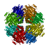
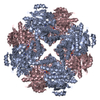
[English] 日本語
Yorodumi
- PDB-4m6u: P. putida mandelate racemase co-crystallized with tartronic acid -

+
Open data


ID or keywords:

Loading...

-
Basic information

Entry
Database: PDB / ID: 4m6u
TitleP. putida mandelate racemase co-crystallized with tartronic acid
ComponentsMandelate racemase
KeywordsISOMERASE / Enolase superfamily enzyme
Function / homology
Function and homology information


mandelate racemase / mandelate racemase activity / mandelate catabolic process / amino acid catabolic process / hydro-lyase activity / metal ion binding
Similarity search - Function
Mandelate racemase / Mandelate racemase / muconate lactonizing enzyme family signature 2. / : / Mandelate racemase / muconate lactonizing enzyme family signature 1. / Mandelate racemase/muconate lactonizing enzyme, conserved site / Mandelate racemase/muconate lactonizing enzyme, N-terminal domain / Mandelate racemase / muconate lactonizing enzyme, N-terminal domain / Mandelate racemase/muconate lactonizing enzyme, C-terminal / Mandelate racemase / muconate lactonizing enzyme, C-terminal domain / Enolase C-terminal domain-like ...Mandelate racemase / Mandelate racemase / muconate lactonizing enzyme family signature 2. / : / Mandelate racemase / muconate lactonizing enzyme family signature 1. / Mandelate racemase/muconate lactonizing enzyme, conserved site / Mandelate racemase/muconate lactonizing enzyme, N-terminal domain / Mandelate racemase / muconate lactonizing enzyme, N-terminal domain / Mandelate racemase/muconate lactonizing enzyme, C-terminal / Mandelate racemase / muconate lactonizing enzyme, C-terminal domain / Enolase C-terminal domain-like / Enolase C-terminal domain-like / Enolase-like C-terminal domain / Enolase-like, N-terminal domain / Enolase-like, N-terminal / Enolase-like, C-terminal domain superfamily / Enolase-like; domain 1 / TIM Barrel / Alpha-Beta Barrel / 2-Layer Sandwich / Alpha Beta
Similarity search - Domain/homology
TARTRONATE / Mandelate racemase
Similarity search - Component
Biological speciesPseudomonas putida (bacteria)
MethodX-RAY DIFFRACTION / SYNCHROTRON / MOLECULAR REPLACEMENT / Resolution: 1.8 Å
AuthorsLietzan, A.D. / St.Maurice, M.
CitationJournal: Biochemistry / Year: 2014
Title: Potent inhibition of mandelate racemase by a fluorinated substrate-product analogue with a novel binding mode.
Authors: Nagar, M. / Lietzan, A.D. / St Maurice, M. / Bearne, S.L.
History
DepositionAug 11, 2013Deposition site: RCSB / Processing site: RCSB
Revision 1.0Mar 5, 2014Provider: repository / Type: Initial release
Revision 1.1Mar 12, 2014Group: Database references
Revision 1.2Jan 24, 2018Group: Structure summary / Category: audit_author / Item: _audit_author.name
Revision 1.3Jan 31, 2018Group: Structure summary / Category: audit_author / Item: _audit_author.name
Revision 1.4Sep 20, 2023Group: Data collection / Database references ...Data collection / Database references / Derived calculations / Refinement description
Category: chem_comp_atom / chem_comp_bond ...chem_comp_atom / chem_comp_bond / database_2 / pdbx_initial_refinement_model / pdbx_struct_conn_angle / struct_conn / struct_ref_seq_dif / struct_site
Item: _database_2.pdbx_DOI / _database_2.pdbx_database_accession ..._database_2.pdbx_DOI / _database_2.pdbx_database_accession / _pdbx_struct_conn_angle.ptnr1_auth_asym_id / _pdbx_struct_conn_angle.ptnr1_auth_comp_id / _pdbx_struct_conn_angle.ptnr1_auth_seq_id / _pdbx_struct_conn_angle.ptnr1_label_asym_id / _pdbx_struct_conn_angle.ptnr1_label_atom_id / _pdbx_struct_conn_angle.ptnr1_label_comp_id / _pdbx_struct_conn_angle.ptnr1_label_seq_id / _pdbx_struct_conn_angle.ptnr2_auth_asym_id / _pdbx_struct_conn_angle.ptnr2_label_asym_id / _pdbx_struct_conn_angle.ptnr3_auth_asym_id / _pdbx_struct_conn_angle.ptnr3_auth_comp_id / _pdbx_struct_conn_angle.ptnr3_auth_seq_id / _pdbx_struct_conn_angle.ptnr3_label_asym_id / _pdbx_struct_conn_angle.ptnr3_label_atom_id / _pdbx_struct_conn_angle.ptnr3_label_comp_id / _pdbx_struct_conn_angle.ptnr3_label_seq_id / _pdbx_struct_conn_angle.value / _struct_conn.pdbx_dist_value / _struct_conn.ptnr1_auth_asym_id / _struct_conn.ptnr1_auth_comp_id / _struct_conn.ptnr1_auth_seq_id / _struct_conn.ptnr1_label_asym_id / _struct_conn.ptnr1_label_atom_id / _struct_conn.ptnr1_label_comp_id / _struct_conn.ptnr1_label_seq_id / _struct_conn.ptnr2_auth_asym_id / _struct_conn.ptnr2_auth_comp_id / _struct_conn.ptnr2_auth_seq_id / _struct_conn.ptnr2_label_asym_id / _struct_conn.ptnr2_label_atom_id / _struct_conn.ptnr2_label_comp_id / _struct_ref_seq_dif.details / _struct_site.pdbx_auth_asym_id / _struct_site.pdbx_auth_comp_id / _struct_site.pdbx_auth_seq_id

-
Structure visualization

Structure viewerMolecule:
MolmilJmol/JSmol

Downloads & links

-
Assembly

Deposited unit
A: Mandelate racemase
B: Mandelate racemase
hetero molecules


Theoretical massNumber of molelcules
Total (without water)82,8946
Polymers82,6092
Non-polymers2854
Water11,476637
1
A: Mandelate racemase
B: Mandelate racemase
hetero molecules

A: Mandelate racemase
B: Mandelate racemase
hetero molecules

A: Mandelate racemase
B: Mandelate racemase
hetero molecules

A: Mandelate racemase
B: Mandelate racemase
hetero molecules


Theoretical massNumber of molelcules
Total (without water)331,57624
Polymers330,4378
Non-polymers1,13916
Water1448
TypeNameSymmetry operationNumber
identity operation1_555x,y,z1
crystal symmetry operation2_555-x,-y,z1
crystal symmetry operation3_555-y,x,z1
crystal symmetry operation4_555y,-x,z1
Buried area29600 Å2
ΔGint-234 kcal/mol
Surface area83400 Å2
MethodPISA
Unit cell
Length a, b, c (Å)149.350, 149.350, 177.752
Angle α, β, γ (deg.)90.00, 90.00, 90.00
Int Tables number97
Space group name H-MI422
Components on special symmetry positions
IDModelComponents
11A-666-

HOH

21A-778-

HOH

-
Components

#1: Protein Mandelate racemase / / MR


Mass: 41304.621 Da / Num. of mol.: 2
Source method: isolated from a genetically manipulated source
Source: (gene. exp.) Pseudomonas putida (bacteria) / Gene: mdlA / Plasmid: pET-52b(+) / Production host: Escherichia coli (E. coli) / Strain (production host): BL21(DE3) / References: UniProt: P11444, mandelate racemase
#2: Chemical ChemComp-MG / MAGNESIUM ION


Mass: 24.305 Da / Num. of mol.: 2 / Source method: obtained synthetically / Formula: Mg
#3: Chemical ChemComp-TTN / TARTRONATE


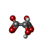
Mass: 118.045 Da / Num. of mol.: 2 / Source method: obtained synthetically / Formula: C3H2O5
#4: Water ChemComp-HOH / water / Water


Mass: 18.015 Da / Num. of mol.: 637 / Source method: isolated from a natural source / Formula: H2O

-
Experimental details

-
Experiment

ExperimentMethod: X-RAY DIFFRACTION / Number of used crystals: 1

-
Sample preparation

CrystalDensity Matthews: 3 Å3/Da / Density % sol: 59 %
Crystal growTemperature: 298 K / Method: vapor diffusion, hanging drop / pH: 8.5
Details: 14% (w/v) PEG 1500, 100 mM glycine, 100 mM Triethanolamine (pH 8.5), VAPOR DIFFUSION, HANGING DROP, temperature 298K

-
Data collection

DiffractionMean temperature: 100 K
Diffraction sourceSource: SYNCHROTRON / Site: APS / Beamline: 21-ID-G / Wavelength: 0.979 Å
DetectorType: MARMOSAIC 300 mm CCD / Detector: CCD / Date: Jun 3, 2013
RadiationMonochromator: C(111) / Protocol: SINGLE WAVELENGTH / Monochromatic (M) / Laue (L): M / Scattering type: x-ray
Radiation wavelengthWavelength: 0.979 Å / Relative weight: 1
ReflectionResolution: 1.8→50 Å / Num. all: 91291 / Num. obs: 90212 / % possible obs: 98.8 % / Observed criterion σ(I): -3 / Redundancy: 10.1 % / Biso Wilson estimate: 13.6 Å2 / Rmerge(I) obs: 0.073 / Net I/σ(I): 29.5
Reflection shellResolution: 1.8→1.83 Å / Redundancy: 8.7 % / Rmerge(I) obs: 0.376 / Mean I/σ(I) obs: 5 / Num. unique all: 4535 / % possible all: 99.8

-
Processing

Software
NameVersionClassification
MD2diffractometer software from EMBLdata collection
PHASERphasing
REFMAC5.7.0032refinement
DENZOdata reduction
SCALEPACKdata scaling
RefinementMethod to determine structure: MOLECULAR REPLACEMENT
Starting model: PDB ENTRY 2MNR
Resolution: 1.8→32.38 Å / Cor.coef. Fo:Fc: 0.97 / Cor.coef. Fo:Fc free: 0.961 / SU B: 1.639 / SU ML: 0.052 / Cross valid method: THROUGHOUT / ESU R: 0.086 / ESU R Free: 0.085 / Stereochemistry target values: MAXIMUM LIKELIHOOD / Details: HYDROGENS HAVE BEEN ADDED IN THE RIDING POSITIONS
RfactorNum. reflection% reflectionSelection details
Rfree0.17073 4570 5 %RANDOM
Rwork0.14662 ---
obs0.14784 86721 98.74 %-
Solvent computationIon probe radii: 0.8 Å / Shrinkage radii: 0.8 Å / VDW probe radii: 1.2 Å / Solvent model: MASK
Displacement parametersBiso mean: 20.043 Å2
Baniso -1Baniso -2Baniso -3
1--0.87 Å20 Å20 Å2
2---0.87 Å20 Å2
3---1.73 Å2
Refinement stepCycle: LAST / Resolution: 1.8→32.38 Å
ProteinNucleic acidLigandSolventTotal
Num. atoms5377 0 18 637 6032
Refine LS restraints
Refine-IDTypeDev idealDev ideal targetNumber
X-RAY DIFFRACTIONr_bond_refined_d0.0170.0195794
X-RAY DIFFRACTIONr_bond_other_d0.0010.025673
X-RAY DIFFRACTIONr_angle_refined_deg1.7541.9717931
X-RAY DIFFRACTIONr_angle_other_deg0.92313046
X-RAY DIFFRACTIONr_dihedral_angle_1_deg6.245774
X-RAY DIFFRACTIONr_dihedral_angle_2_deg39.06324.027221
X-RAY DIFFRACTIONr_dihedral_angle_3_deg13.00215941
X-RAY DIFFRACTIONr_dihedral_angle_4_deg17.7061532
X-RAY DIFFRACTIONr_chiral_restr0.1220.2933
X-RAY DIFFRACTIONr_gen_planes_refined0.0090.0216613
X-RAY DIFFRACTIONr_gen_planes_other0.0030.021251
X-RAY DIFFRACTIONr_nbd_refined
X-RAY DIFFRACTIONr_nbd_other
X-RAY DIFFRACTIONr_nbtor_refined
X-RAY DIFFRACTIONr_nbtor_other
X-RAY DIFFRACTIONr_xyhbond_nbd_refined
X-RAY DIFFRACTIONr_xyhbond_nbd_other
X-RAY DIFFRACTIONr_metal_ion_refined
X-RAY DIFFRACTIONr_metal_ion_other
X-RAY DIFFRACTIONr_symmetry_vdw_refined
X-RAY DIFFRACTIONr_symmetry_vdw_other
X-RAY DIFFRACTIONr_symmetry_hbond_refined
X-RAY DIFFRACTIONr_symmetry_hbond_other
X-RAY DIFFRACTIONr_symmetry_metal_ion_refined
X-RAY DIFFRACTIONr_symmetry_metal_ion_other
X-RAY DIFFRACTIONr_mcbond_it1.6161.8522992
X-RAY DIFFRACTIONr_mcbond_other1.6161.8512991
X-RAY DIFFRACTIONr_mcangle_it2.3832.7723749
X-RAY DIFFRACTIONr_mcangle_other2.3822.7733750
X-RAY DIFFRACTIONr_scbond_it2.1412.0312802
X-RAY DIFFRACTIONr_scbond_other2.1382.032799
X-RAY DIFFRACTIONr_scangle_it
X-RAY DIFFRACTIONr_scangle_other3.182.9814163
X-RAY DIFFRACTIONr_long_range_B_refined4.92515.4026805
X-RAY DIFFRACTIONr_long_range_B_other4.55814.7396454
X-RAY DIFFRACTIONr_rigid_bond_restr
X-RAY DIFFRACTIONr_sphericity_free
X-RAY DIFFRACTIONr_sphericity_bonded
LS refinement shellResolution: 1.8→1.847 Å / Total num. of bins used: 20
RfactorNum. reflection% reflection
Rfree0.264 313 -
Rwork0.2 6393 -
obs-6393 99.23 %

+
About Yorodumi

-
News

-
Feb 9, 2022. New format data for meta-information of EMDB entries

New format data for meta-information of EMDB entries

  • Version 3 of the EMDB header file is now the official format.
  • The previous official version 1.9 will be removed from the archive.

Related info.:EMDB header

External links:wwPDB to switch to version 3 of the EMDB data model

-
Aug 12, 2020. Covid-19 info

Covid-19 info

URL: https://pdbj.org/emnavi/covid19.php

New page: Covid-19 featured information page in EM Navigator.

Related info.:Covid-19 info / Mar 5, 2020. Novel coronavirus structure data

+
Mar 5, 2020. Novel coronavirus structure data

Novel coronavirus structure data

Related info.:Yorodumi Speices / Aug 12, 2020. Covid-19 info

External links:COVID-19 featured content - PDBj / Molecule of the Month (242):Coronavirus Proteases

+
Jan 31, 2019. EMDB accession codes are about to change! (news from PDBe EMDB page)

EMDB accession codes are about to change! (news from PDBe EMDB page)

  • The allocation of 4 digits for EMDB accession codes will soon come to an end. Whilst these codes will remain in use, new EMDB accession codes will include an additional digit and will expand incrementally as the available range of codes is exhausted. The current 4-digit format prefixed with “EMD-” (i.e. EMD-XXXX) will advance to a 5-digit format (i.e. EMD-XXXXX), and so on. It is currently estimated that the 4-digit codes will be depleted around Spring 2019, at which point the 5-digit format will come into force.
  • The EM Navigator/Yorodumi systems omit the EMD- prefix.

Related info.:Q: What is EMD? / ID/Accession-code notation in Yorodumi/EM Navigator

External links:EMDB Accession Codes are Changing Soon! / Contact to PDBj

+
Jul 12, 2017. Major update of PDB

Major update of PDB

  • wwPDB released updated PDB data conforming to the new PDBx/mmCIF dictionary.
  • This is a major update changing the version number from 4 to 5, and with Remediation, in which all the entries are updated.
  • In this update, many items about electron microscopy experimental information are reorganized (e.g. em_software).
  • Now, EM Navigator and Yorodumi are based on the updated data.

External links:wwPDB Remediation / Enriched Model Files Conforming to OneDep Data Standards Now Available in the PDB FTP Archive

-
Yorodumi

Thousand views of thousand structures

  • Yorodumi is a browser for structure data from EMDB, PDB, SASBDB, etc.
  • This page is also the successor to EM Navigator detail page, and also detail information page/front-end page for Omokage search.
  • The word "yorodu" (or yorozu) is an old Japanese word meaning "ten thousand". "mi" (miru) is to see.

Related info.:EMDB / PDB / SASBDB / Comparison of 3 databanks / Yorodumi Search / Aug 31, 2016. New EM Navigator & Yorodumi / Yorodumi Papers / Jmol/JSmol / Function and homology information / Changes in new EM Navigator and Yorodumi

Read more