 Movie
Movie Controller

[English] 日本語
 Yorodumi
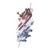
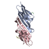
- PDB-4m5s: Human alphaB crystallin core domain in complex with C-terminal peptide -
+ Open data

- Basic information
| Entry | Database: PDB / ID: 4m5s | ||||||
|---|---|---|---|---|---|---|---|
| Title | Human alphaB crystallin core domain in complex with C-terminal peptide | ||||||
 Components Components | (Alpha-crystallin B chain CRYAB) x 2 CRYAB) x 2 | ||||||
 Keywords Keywords | CHAPERONE / small heat shock protein / amyloid | ||||||
| Function / homology |  Function and homology information Function and homology informationmicrotubule polymerization or depolymerization / negative regulation of intracellular transport / apoptotic process involved in morphogenesis / cardiac myofibril / regulation of programmed cell death / tubulin complex assembly / structural constituent of eye lens / negative regulation of amyloid fibril formation / M band / lens development in camera-type eye ...microtubule polymerization or depolymerization / negative regulation of intracellular transport / apoptotic process involved in morphogenesis / cardiac myofibril / regulation of programmed cell death / tubulin complex assembly / structural constituent of eye lens / negative regulation of amyloid fibril formation / M band / lens development in camera-type eye / muscle organ development / actin filament bundle / HSF1-dependent transactivation / negative regulation of reactive oxygen species metabolic process / negative regulation of protein-containing complex assembly / stress-activated MAPK cascade / muscle contraction / synaptic membrane / response to hydrogen peroxide / cellular response to gamma radiation / negative regulation of cell growth / Z disc / unfolded protein binding / protein folding / response to estradiol / amyloid-beta binding / response to heat / perikaryon / protein refolding / microtubule binding / dendritic spine / lysosome / response to hypoxia / protein stabilization / axon / negative regulation of gene expression / negative regulation of DNA-templated transcription / protein-containing complex binding / negative regulation of apoptotic process / structural molecule activity / cell surface / protein homodimerization activity / protein-containing complex / mitochondrion / extracellular exosome / nucleoplasm / identical protein binding / metal ion binding / nucleus / cytosol / cytoplasmSimilarity search - Function | ||||||
| Biological species |  Homo sapiens (human) Homo sapiens (human) | ||||||
| Method | X-RAY DIFFRACTION / SYNCHROTRON / MOLECULAR REPLACEMENT / Resolution: 1.37 Å | ||||||
 Authors Authors | Laganowsky, A. / Cascio, D. / Sawaya, M.R. / Eisenberg, D. | ||||||
 Citation Citation | Journal: Proc.Natl.Acad.Sci.USA / Year: 2014 Title: The structured core domain of alpha B-crystallin can prevent amyloid fibrillation and associated toxicity. Authors: Hochberg, G.K. / Ecroyd, H. / Liu, C. / Cox, D. / Cascio, D. / Sawaya, M.R. / Collier, M.P. / Stroud, J. / Carver, J.A. / Baldwin, A.J. / Robinson, C.V. / Eisenberg, D.S. / Benesch, J.L. / Laganowsky, A. | ||||||
| History |
|
- Structure visualization
| Structure viewer | Molecule: MolmilJmol/JSmol |
|---|
- Downloads & links
-Download
| PDBx/mmCIF format | 4m5s.cif.gz | 58.3 KB | Display | PDBx/mmCIF format |
|---|---|---|---|---|
| PDB format | pdb4m5s.ent.gz | 42.4 KB | Display | PDB format |
| PDBx/mmJSON format | 4m5s.json.gz | Tree view | PDBx/mmJSON format | |
| Others |  Other downloads Other downloads |
-Validation report
| Arichive directory | https://data.pdbj.org/pub/pdb/validation_reports/m5/4m5sftp://data.pdbj.org/pub/pdb/validation_reports/m5/4m5s | HTTPS FTP |
|---|
-Related structure data


-Links
 PDBj
PDBj


- Assembly
| Deposited unit | 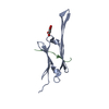
| ||||||||
|---|---|---|---|---|---|---|---|---|---|
| 1 | 
| ||||||||
| 2 |
| ||||||||
| Unit cell |
|
-Components
| #1: Protein | CRYAB / Alpha(B)-crystallin / Heat shock protein beta-5 / HspB5 / Renal carcinoma antigen NY-REN-27 / ...Alpha(B)-crystallin / Heat shock protein beta-5 / HspB5 / Renal carcinoma antigen NY-REN-27 / Rosenthal fiber component Mass: 9957.241 Da / Num. of mol.: 1 / Fragment: core domain (UNP residues 68-153) Source method: isolated from a genetically manipulated source Source: (gene. exp.) Homo sapiens (human) / Gene: CRYA2, CRYAB / Plasmid: pET28 / Production host:  Escherichia coli (E. coli) / Strain (production host): BL21(DE3) Rosetta 2 / References: UniProt: P02511 Escherichia coli (E. coli) / Strain (production host): BL21(DE3) Rosetta 2 / References: UniProt: P02511 |
|---|---|
| #2: Protein/peptide | CRYAB / Alpha(B)-crystallin / Heat shock protein beta-5 / HspB5 / Renal carcinoma antigen NY-REN-27 / ...Alpha(B)-crystallin / Heat shock protein beta-5 / HspB5 / Renal carcinoma antigen NY-REN-27 / Rosenthal fiber component Mass: 1173.320 Da / Num. of mol.: 1 / Fragment: C-terminal peptide (UNP residues 156-164) Source method: isolated from a genetically manipulated source Source: (gene. exp.) Homo sapiens (human) / Gene: CRYA2, CRYAB / Plasmid: pET15-MBP / Production host: Escherichia coli (E. coli) / Strain (production host): BL21(DE3) / References: UniProt: P02511 |
| #3: Chemical | ChemComp-SIN / Succinic acid  Mass: 118.088 Da / Num. of mol.: 1 / Source method: obtained synthetically / Formula: C4H6O4 Mass: 118.088 Da / Num. of mol.: 1 / Source method: obtained synthetically / Formula: C4H6O4 |
| #4: Water | ChemComp-HOH / Water Mass: 18.015 Da / Num. of mol.: 70 / Source method: isolated from a natural source / Formula: H2O Mass: 18.015 Da / Num. of mol.: 70 / Source method: isolated from a natural source / Formula: H2O |
-Experimental details
-Experiment
| Experiment | Method: X-RAY DIFFRACTION / Number of used crystals: 1 |
|---|
- Sample preparation
| Crystal | Density Matthews: 2.34 Å3/Da / Density % sol: 47.34 % |
|---|---|
| Crystal grow | Temperature: 298 K / Method: vapor diffusion, hanging drop / pH: 6 Details: 0.1 M SPG, pH 6.0, 25% PEG1500, VAPOR DIFFUSION, HANGING DROP, temperature 298K |
-Data collection
| Diffraction | Mean temperature: 100 K | |||||||||||||||||||||||||||||||||||||||||||||||||||||||||||||||||||||||||||||||||||||||||||||||||||||||||||||||||||||||||||||||||||||||||||||||||||
|---|---|---|---|---|---|---|---|---|---|---|---|---|---|---|---|---|---|---|---|---|---|---|---|---|---|---|---|---|---|---|---|---|---|---|---|---|---|---|---|---|---|---|---|---|---|---|---|---|---|---|---|---|---|---|---|---|---|---|---|---|---|---|---|---|---|---|---|---|---|---|---|---|---|---|---|---|---|---|---|---|---|---|---|---|---|---|---|---|---|---|---|---|---|---|---|---|---|---|---|---|---|---|---|---|---|---|---|---|---|---|---|---|---|---|---|---|---|---|---|---|---|---|---|---|---|---|---|---|---|---|---|---|---|---|---|---|---|---|---|---|---|---|---|---|---|---|---|---|
| Diffraction source | Source: SYNCHROTRON / Site: APS  / Beamline: 24-ID-C / Wavelength: 0.9794 Å / Beamline: 24-ID-C / Wavelength: 0.9794 Å | |||||||||||||||||||||||||||||||||||||||||||||||||||||||||||||||||||||||||||||||||||||||||||||||||||||||||||||||||||||||||||||||||||||||||||||||||||
| Detector | Type: ADSC QUANTUM 315 / Detector: CCD / Date: Dec 10, 2010 | |||||||||||||||||||||||||||||||||||||||||||||||||||||||||||||||||||||||||||||||||||||||||||||||||||||||||||||||||||||||||||||||||||||||||||||||||||
| Radiation | Monochromator: Cryo-Cooled double crystal Si(111) / Protocol: SINGLE WAVELENGTH / Monochromatic (M) / Laue (L): M / Scattering type: x-ray | |||||||||||||||||||||||||||||||||||||||||||||||||||||||||||||||||||||||||||||||||||||||||||||||||||||||||||||||||||||||||||||||||||||||||||||||||||
| Radiation wavelength | Wavelength: 0.9794 Å / Relative weight: 1 | |||||||||||||||||||||||||||||||||||||||||||||||||||||||||||||||||||||||||||||||||||||||||||||||||||||||||||||||||||||||||||||||||||||||||||||||||||
| Reflection | Resolution: 1.37→19.569 Å / Num. obs: 21884 / % possible obs: 97.5 % / Observed criterion σ(I): -3 / Biso Wilson estimate: 15.64 Å2 / Rmerge(I) obs: 0.042 / Net I/σ(I): 25.13 | |||||||||||||||||||||||||||||||||||||||||||||||||||||||||||||||||||||||||||||||||||||||||||||||||||||||||||||||||||||||||||||||||||||||||||||||||||
| Reflection shell | Diffraction-ID: 1
|
- Processing
| Software |
| |||||||||||||||||||||||||||||||||||||||||||||||||||||||||||||||
|---|---|---|---|---|---|---|---|---|---|---|---|---|---|---|---|---|---|---|---|---|---|---|---|---|---|---|---|---|---|---|---|---|---|---|---|---|---|---|---|---|---|---|---|---|---|---|---|---|---|---|---|---|---|---|---|---|---|---|---|---|---|---|---|---|
| Refinement | Method to determine structure: MOLECULAR REPLACEMENT / Resolution: 1.37→19.569 Å / Occupancy max: 1 / Occupancy min: 0.24 / FOM work R set: 0.8864 / SU ML: 0.12 / σ(F): 1.99 / Phase error: 17.92 / Stereochemistry target values: ML
| |||||||||||||||||||||||||||||||||||||||||||||||||||||||||||||||
| Solvent computation | Shrinkage radii: 0.9 Å / VDW probe radii: 1.11 Å / Solvent model: FLAT BULK SOLVENT MODEL | |||||||||||||||||||||||||||||||||||||||||||||||||||||||||||||||
| Displacement parameters | Biso max: 77.51 Å2 / Biso mean: 24.1899 Å2 / Biso min: 8.13 Å2 | |||||||||||||||||||||||||||||||||||||||||||||||||||||||||||||||
| Refinement step | Cycle: LAST / Resolution: 1.37→19.569 Å
| |||||||||||||||||||||||||||||||||||||||||||||||||||||||||||||||
| Refine LS restraints |
| |||||||||||||||||||||||||||||||||||||||||||||||||||||||||||||||
| LS refinement shell | Refine-ID: X-RAY DIFFRACTION / Total num. of bins used: 8
|
