 Movie
Movie Controller

[English] 日本語
 Yorodumi
- PDB-4lz0: A236G Epi-isozizaene synthase: Complex with Mg, inorganic pyropho... -
+ Open data

- Basic information
| Entry | Database: PDB / ID: 4lz0 | ||||||
|---|---|---|---|---|---|---|---|
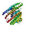
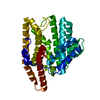
| Title | A236G Epi-isozizaene synthase: Complex with Mg, inorganic pyrophosphate and benzyl triethyl ammonium cation | ||||||
 Components Components | Epi-isozizaene synthase | ||||||
 Keywords Keywords | LYASE / Class I terpene cyclase | ||||||
| Function / homology |  Function and homology informationepi-isozizaene synthase / epi-isozizaene synthase activity / terpene synthase activity / metal ion binding Function and homology informationepi-isozizaene synthase / epi-isozizaene synthase activity / terpene synthase activity / metal ion bindingSimilarity search - Function | ||||||
| Biological species |  Streptomyces coelicolor (bacteria) Streptomyces coelicolor (bacteria) | ||||||
| Method | X-RAY DIFFRACTION / SYNCHROTRON / MOLECULAR REPLACEMENT / Resolution: 1.754 Å | ||||||
 Authors Authors | Li, R. / Chou, W. / Himmelberger, J.A. / Litwin, K. / Harris, G. / Cane, D.E. / Christianson, D.W. | ||||||
 Citation Citation | Journal: Biochemistry / Year: 2014 Title: Reprogramming the Chemodiversity of Terpenoid Cyclization by Remolding the Active Site Contour of epi-Isozizaene Synthase. Authors: Li, R. / Chou, W.K. / Himmelberger, J.A. / Litwin, K.M. / Harris, G.G. / Cane, D.E. / Christianson, D.W. | ||||||
| History |
|
- Structure visualization
| Structure viewer | Molecule: MolmilJmol/JSmol |
|---|
- Downloads & links
-Download
| PDBx/mmCIF format | 4lz0.cif.gz | 93 KB | Display | PDBx/mmCIF format |
|---|---|---|---|---|
| PDB format | pdb4lz0.ent.gz | 67.9 KB | Display | PDB format |
| PDBx/mmJSON format | 4lz0.json.gz | Tree view | PDBx/mmJSON format | |
| Others |  Other downloads Other downloads |
-Validation report
| Arichive directory | https://data.pdbj.org/pub/pdb/validation_reports/lz/4lz0ftp://data.pdbj.org/pub/pdb/validation_reports/lz/4lz0 | HTTPS FTP |
|---|
-Related structure data
| Related structure data | 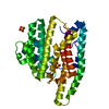 4ltvC 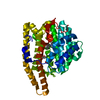 4ltzC  4luuC  4lxwC  4lz3C  4lzcC  3kb9S S: Starting model for refinement C: citing same article ( |
|---|---|
| Similar structure data |


-Links
 PDBj
PDBj


- Assembly
| Deposited unit | 
| ||||||||
|---|---|---|---|---|---|---|---|---|---|
| 1 |
| ||||||||
| Unit cell |
|
-Components
-Protein , 1 types, 1 molecules A
| #1: Protein | / EIZS / Sesquiterpene cyclase / Sesquiterpene synthase Mass: 43701.984 Da / Num. of mol.: 1 / Mutation: A236G Source method: isolated from a genetically manipulated source Source: (gene. exp.) Streptomyces coelicolor (bacteria) / Strain: A3(2) / Gene: cyc1, SCO5222, SC7E4.19 / Plasmid: pET28a(+) / Production host: Escherichia coli (E. coli) / Strain (production host): BL21(DE3) / References: UniProt: Q9K499, epi-isozizaene synthase |
|---|
-Non-polymers , 5 types, 289 molecules 


| #2: Chemical |  Mass: 24.305 Da / Num. of mol.: 3 / Source method: obtained synthetically / Formula: Mg Mass: 24.305 Da / Num. of mol.: 3 / Source method: obtained synthetically / Formula: Mg#3: Chemical | ChemComp-POP / | Pyrophosphate Mass: 175.959 Da / Num. of mol.: 1 / Source method: obtained synthetically / Formula: H2O7P2 Mass: 175.959 Da / Num. of mol.: 1 / Source method: obtained synthetically / Formula: H2O7P2#4: Chemical | ChemComp-BTM / |  Mass: 192.320 Da / Num. of mol.: 1 / Source method: obtained synthetically / Formula: C13H22N Mass: 192.320 Da / Num. of mol.: 1 / Source method: obtained synthetically / Formula: C13H22N#5: Chemical | ChemComp-SO4 / | Sulfate Mass: 96.063 Da / Num. of mol.: 1 / Source method: obtained synthetically / Formula: SO4 Mass: 96.063 Da / Num. of mol.: 1 / Source method: obtained synthetically / Formula: SO4#6: Water | ChemComp-HOH / | WaterMass: 18.015 Da / Num. of mol.: 283 / Source method: isolated from a natural source / Formula: H2O |
|---|
-Experimental details
-Experiment
| Experiment | Method: X-RAY DIFFRACTION / Number of used crystals: 1 |
|---|
- Sample preparation
| Crystal | Density Matthews: 2.15 Å3/Da / Density % sol: 42.77 % |
|---|---|
| Crystal grow | Temperature: 298 K / Method: vapor diffusion, hanging drop / pH: 5.5 Details: a 4 uL drop of protein solution [8 mg/mL A236G EIZS, 20 mM Tris-HCl (pH 7.5), 300 mM NaCl, 10 mM MgCl2, 10% glycerol, 1 mM TCEP, 2 mM sodium pyrophosphate, 2 mM benzyltriethylammonium ...Details: a 4 uL drop of protein solution [8 mg/mL A236G EIZS, 20 mM Tris-HCl (pH 7.5), 300 mM NaCl, 10 mM MgCl2, 10% glycerol, 1 mM TCEP, 2 mM sodium pyrophosphate, 2 mM benzyltriethylammonium chloride (BTAC)] was added to a 4 uL drop of precipitant solution [100 mM Bis-Tris (pH 5.5), 25-28% polyethylene glycol 3350, 0.2 M (NH4)2SO4] and equilibrated against a 1 mL reservoir of precipitant solution at 298K., VAPOR DIFFUSION, HANGING DROP |
-Data collection
| Diffraction | Mean temperature: 100 K |
|---|---|
| Diffraction source | Source: SYNCHROTRON / Site: APS  / Beamline: 24-ID-C / Wavelength: 0.9795 Å / Beamline: 24-ID-C / Wavelength: 0.9795 Å |
| Detector | Type: ADSC QUANTUM 315 / Detector: CCD / Date: Dec 10, 2009 / Details: Kirpatrick Baez focusing mirrors |
| Radiation | Monochromator: Si(111) double crystal monochromator / Protocol: SINGLE WAVELENGTH / Monochromatic (M) / Laue (L): M / Scattering type: x-ray |
| Radiation wavelength | Wavelength: 0.9795 Å / Relative weight: 1 |
| Reflection | Resolution: 1.754→50 Å / Num. obs: 37050 / % possible obs: 99.2 % / Observed criterion σ(F): 0 / Observed criterion σ(I): -3 / Redundancy: 3.3 % / Rmerge(I) obs: 0.071 / Rsym value: 0.071 / Net I/σ(I): 13.689 |
| Reflection shell | Resolution: 1.76→1.82 Å / Redundancy: 3.1 % / Rmerge(I) obs: 0.339 / Mean I/σ(I) obs: 2.73 / Rsym value: 0.339 / % possible all: 98.2 |
- Processing
| Software |
| |||||||||||||||||||||||||||||||||||||||||||||||||||||||||||||||||||||||||||||||||||||||||||||||||||||||||
|---|---|---|---|---|---|---|---|---|---|---|---|---|---|---|---|---|---|---|---|---|---|---|---|---|---|---|---|---|---|---|---|---|---|---|---|---|---|---|---|---|---|---|---|---|---|---|---|---|---|---|---|---|---|---|---|---|---|---|---|---|---|---|---|---|---|---|---|---|---|---|---|---|---|---|---|---|---|---|---|---|---|---|---|---|---|---|---|---|---|---|---|---|---|---|---|---|---|---|---|---|---|---|---|---|---|---|
| Refinement | Method to determine structure: MOLECULAR REPLACEMENT Starting model: PDBID: 3KB9 Resolution: 1.754→37.431 Å / SU ML: 0.18 / σ(F): 1.34 / Phase error: 19.68 / Stereochemistry target values: ML
| |||||||||||||||||||||||||||||||||||||||||||||||||||||||||||||||||||||||||||||||||||||||||||||||||||||||||
| Solvent computation | Shrinkage radii: 0.9 Å / VDW probe radii: 1.11 Å / Solvent model: FLAT BULK SOLVENT MODEL | |||||||||||||||||||||||||||||||||||||||||||||||||||||||||||||||||||||||||||||||||||||||||||||||||||||||||
| Displacement parameters |
| |||||||||||||||||||||||||||||||||||||||||||||||||||||||||||||||||||||||||||||||||||||||||||||||||||||||||
| Refinement step | Cycle: LAST / Resolution: 1.754→37.431 Å
| |||||||||||||||||||||||||||||||||||||||||||||||||||||||||||||||||||||||||||||||||||||||||||||||||||||||||
| Refine LS restraints |
| |||||||||||||||||||||||||||||||||||||||||||||||||||||||||||||||||||||||||||||||||||||||||||||||||||||||||
| LS refinement shell |
|
