 Movie
Movie Controller


+ Open data

- Basic information
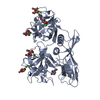
| Entry | Database: PDB / ID: 4jut | ||||||
|---|---|---|---|---|---|---|---|
| Title | Crystal structure of a mutant fragment of Human HSPB6 | ||||||
 Components Components | Heat shock protein beta-6 Heat shock response Heat shock response | ||||||
 Keywords Keywords | CHAPERONE / Small heat shock protein / alpha-crystallin domain | ||||||
| Function / homology |  Function and homology information Function and homology informationstructural constituent of eye lens / chaperone-mediated protein folding / protein folding chaperone / positive regulation of angiogenesis / unfolded protein binding / response to heat / protein-folding chaperone binding / protein refolding / nuclear speck / negative regulation of apoptotic process ...structural constituent of eye lens / chaperone-mediated protein folding / protein folding chaperone / positive regulation of angiogenesis / unfolded protein binding / response to heat / protein-folding chaperone binding / protein refolding / nuclear speck / negative regulation of apoptotic process / protein homodimerization activity / mitochondrion / extracellular region / nucleus / cytosol / cytoplasmSimilarity search - Function | ||||||
| Biological species |  Homo sapiens (human) Homo sapiens (human) | ||||||
| Method | X-RAY DIFFRACTION / SYNCHROTRON / MOLECULAR REPLACEMENT / molecular replacement / Resolution: 2.196 Å | ||||||
 Authors Authors | Weeks, S.D. / Baranova, E.V. / Beelen, S. / Heirbaut, M. / Gusev, N.B. / Strelkov, S.V. | ||||||
 Citation Citation | Journal: J.Struct.Biol. / Year: 2014 Title: Molecular structure and dynamics of the dimeric human small heat shock protein HSPB6. Authors: Weeks, S.D. / Baranova, E.V. / Heirbaut, M. / Beelen, S. / Shkumatov, A.V. / Gusev, N.B. / Strelkov, S.V. | ||||||
| History |
|
- Structure visualization
| Structure viewer | Molecule: MolmilJmol/JSmol |
|---|
- Downloads & links
-Download
| PDBx/mmCIF format | 4jut.cif.gz | 142.3 KB | Display | PDBx/mmCIF format |
|---|---|---|---|---|
| PDB format | pdb4jut.ent.gz | 115.7 KB | Display | PDB format |
| PDBx/mmJSON format | 4jut.json.gz | Tree view | PDBx/mmJSON format | |
| Others |  Other downloads Other downloads |
-Validation report
| Arichive directory | https://data.pdbj.org/pub/pdb/validation_reports/ju/4jutftp://data.pdbj.org/pub/pdb/validation_reports/ju/4jut | HTTPS FTP |
|---|
-Related structure data


-Links
 PDBj
PDBj


- Assembly
| Deposited unit | 
| ||||||||
|---|---|---|---|---|---|---|---|---|---|
| 1 | 
| ||||||||
| 2 | 
| ||||||||
| Unit cell |
|
-Components
| #1: Protein | Heat shock response / HspB6 / Heat shock 20 kDa-like protein p20 Mass: 11117.516 Da / Num. of mol.: 8 / Fragment: UNP residues 57-160 / Mutation: E51A, E52A Source method: isolated from a genetically manipulated source Source: (gene. exp.) Homo sapiens (human) / Gene: HSPB6 / Plasmid: pPEPTEV / Production host:  Escherichia coli (E. coli) / Strain (production host): BL21(DE3) / References: UniProt: O14558 Escherichia coli (E. coli) / Strain (production host): BL21(DE3) / References: UniProt: O14558#2: Chemical | ChemComp-GOL / Glycerol  Mass: 92.094 Da / Num. of mol.: 6 / Source method: obtained synthetically / Formula: C3H8O3 Mass: 92.094 Da / Num. of mol.: 6 / Source method: obtained synthetically / Formula: C3H8O3#3: Water | ChemComp-HOH / | Water Mass: 18.015 Da / Num. of mol.: 176 / Source method: isolated from a natural source / Formula: H2O Mass: 18.015 Da / Num. of mol.: 176 / Source method: isolated from a natural source / Formula: H2O |
|---|
-Experimental details
-Experiment
| Experiment | Method: X-RAY DIFFRACTION / Number of used crystals: 1 |
|---|
- Sample preparation
| Crystal | Density Matthews: 2.78 Å3/Da / Density % sol: 55.71 % |
|---|---|
| Crystal grow | Temperature: 277 K / Method: hanging drop vapor diffusion / pH: 7.5 Details: 0.1M HEPES (pH 7.5), 0.2M litium nitrate, 20% PEG 3350, hanging drop vapor diffusion, temperature 277K |
-Data collection
| Diffraction | Mean temperature: 100 K |
|---|---|
| Diffraction source | Source: SYNCHROTRON / Site: SLS  / Beamline: X06DA / Wavelength: 0.99 Å / Beamline: X06DA / Wavelength: 0.99 Å |
| Detector | Type: MARMOSAIC 225 mm CCD / Detector: CCD / Date: Jul 12, 2008 |
| Radiation | Monochromator: Kirkpatrick-Baez pair of bi-morph mirrors plus channel cut cryogenically cooled monochromator crystal Protocol: SINGLE WAVELENGTH / Monochromatic (M) / Laue (L): M / Scattering type: x-ray |
| Radiation wavelength | Wavelength: 0.99 Å / Relative weight: 1 |
| Reflection | Resolution: 2.196→43.01 Å / Num. all: 49462 / Num. obs: 49462 / % possible obs: 99.4 % / Observed criterion σ(F): 2 / Observed criterion σ(I): 2 / Redundancy: 3.8 % / Biso Wilson estimate: 38.7 Å2 / Rsym value: 0.077 |
-Phasing
| Phasing | Method: molecular replacement |
|---|
- Processing
| Software |
| |||||||||||||||||||||||||||||||||||||||||||||||||||||||||||||||||||||||||||||||||||||||||||||||||||||||||||||||||||||||||||||||||||||
|---|---|---|---|---|---|---|---|---|---|---|---|---|---|---|---|---|---|---|---|---|---|---|---|---|---|---|---|---|---|---|---|---|---|---|---|---|---|---|---|---|---|---|---|---|---|---|---|---|---|---|---|---|---|---|---|---|---|---|---|---|---|---|---|---|---|---|---|---|---|---|---|---|---|---|---|---|---|---|---|---|---|---|---|---|---|---|---|---|---|---|---|---|---|---|---|---|---|---|---|---|---|---|---|---|---|---|---|---|---|---|---|---|---|---|---|---|---|---|---|---|---|---|---|---|---|---|---|---|---|---|---|---|---|---|
| Refinement | Method to determine structure: MOLECULAR REPLACEMENT / Resolution: 2.196→43.005 Å / Occupancy max: 1 / Occupancy min: 0.36 / SU ML: 0.38 / σ(F): 1.35 / Phase error: 26.57 / Stereochemistry target values: ML
| |||||||||||||||||||||||||||||||||||||||||||||||||||||||||||||||||||||||||||||||||||||||||||||||||||||||||||||||||||||||||||||||||||||
| Solvent computation | Shrinkage radii: 0.86 Å / VDW probe radii: 1.1 Å / Solvent model: FLAT BULK SOLVENT MODEL / Bsol: 38.102 Å2 / ksol: 0.338 e/Å3 | |||||||||||||||||||||||||||||||||||||||||||||||||||||||||||||||||||||||||||||||||||||||||||||||||||||||||||||||||||||||||||||||||||||
| Displacement parameters | Biso max: 127.93 Å2 / Biso mean: 43.7462 Å2 / Biso min: 8.38 Å2
| |||||||||||||||||||||||||||||||||||||||||||||||||||||||||||||||||||||||||||||||||||||||||||||||||||||||||||||||||||||||||||||||||||||
| Refinement step | Cycle: LAST / Resolution: 2.196→43.005 Å
| |||||||||||||||||||||||||||||||||||||||||||||||||||||||||||||||||||||||||||||||||||||||||||||||||||||||||||||||||||||||||||||||||||||
| Refine LS restraints |
| |||||||||||||||||||||||||||||||||||||||||||||||||||||||||||||||||||||||||||||||||||||||||||||||||||||||||||||||||||||||||||||||||||||
| LS refinement shell | Refine-ID: X-RAY DIFFRACTION / Total num. of bins used: 18
|
