Entry Database : PDB / ID : 4h58Title BRAF in complex with compound 3 Serine/threonine-protein kinase B-raf Keywords / / / Function / homology Function Domain/homology Component
/ / / / / / / / / / / / / / / / / / / / / / / / / / / / / / / / / / / / / / / / / / / / / / / / / / / / / / / / / / / / / / / / / / / / / / / / / / / / / / / / / / / / / / / / / / / / / / / / / / / / / / / / / / / / / / / / / / / / / / / / / / / / / / Biological species Homo sapiens (human)Method / / / Resolution : 3.1 Å Authors Vasbinder, M. / Aquila, B. / Augustin, M. / Chueng, T. / Cook, D. / Drew, L. / Fauber, B. / Glossop, S. / Godin, R. / Grondine, M. ...Vasbinder, M. / Aquila, B. / Augustin, M. / Chueng, T. / Cook, D. / Drew, L. / Fauber, B. / Glossop, S. / Godin, R. / Grondine, M. / Hennessy, E. / Johannes, J. / Lee, S. / Lyne, P. / Moertl, M. / Omer, C. / Palakurthi, S. / Pontz, T. / Read, J. / Sha, L. / Shen, M. / Steinbacher, S. / Wang, H. / Wu, A. / Ye, M. / Bagal, B. Journal : J.Med.Chem. / Year : 2013Title : Discovery and Optimization of a Novel Series of Potent Mutant B-Raf(V600E) Selective Kinase Inhibitors.Authors: Vasbinder, M.M. / Aquila, B. / Augustin, M. / Chen, H. / Cheung, T. / Cook, D. / Drew, L. / Fauber, B.P. / Glossop, S. / Grondine, M. / Hennessy, E. / Johannes, J. / Lee, S. / Lyne, P. / ... Authors : Vasbinder, M.M. / Aquila, B. / Augustin, M. / Chen, H. / Cheung, T. / Cook, D. / Drew, L. / Fauber, B.P. / Glossop, S. / Grondine, M. / Hennessy, E. / Johannes, J. / Lee, S. / Lyne, P. / Mortl, M. / Omer, C. / Palakurthi, S. / Pontz, T. / Read, J. / Sha, L. / Shen, M. / Steinbacher, S. / Wang, H. / Wu, A. / Ye, M. History Deposition Sep 18, 2012 Deposition site / Processing site Revision 1.0 Feb 27, 2013 Provider / Type Revision 1.1 Mar 27, 2013 Group Revision 1.2 Nov 15, 2017 Group / Category Revision 1.3 Sep 20, 2023 Group Data collection / Database references ... Data collection / Database references / Derived calculations / Refinement description Category chem_comp_atom / chem_comp_bond ... chem_comp_atom / chem_comp_bond / database_2 / pdbx_initial_refinement_model / struct_site Item _database_2.pdbx_DOI / _database_2.pdbx_database_accession ... _database_2.pdbx_DOI / _database_2.pdbx_database_accession / _struct_site.pdbx_auth_asym_id / _struct_site.pdbx_auth_comp_id / _struct_site.pdbx_auth_seq_id
Show all Show less
 Movie
Movie Controller


 Open data
 Basic information
 Components
 Keywords
 protein kinase / structure based drug discovery / TRANSFERASE-TRANSFERASE INHIBITOR complex
 Function and homology information
 Authors
 Citation
 Structure visualization
 Downloads & links
 Other downloads
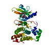


 PDBj
PDBj


 Assembly


 Mass: 385.462 Da / Num. of mol.: 1 / Source method: obtained synthetically / Formula: C23H23N5O
 Mass: 35.453 Da / Num. of mol.: 1 / Source method: obtained synthetically / Formula: Cl
 Mass: 18.015 Da / Num. of mol.: 15 / Source method: isolated from a natural source / Formula: H2O
 Sample preparation
 / Beamline: X06SA / Wavelength: 0.9999 Å
 Processing