| 登録情報 | データベース: PDB / ID: 4gk2
|
|---|
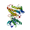
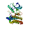
| タイトル | Human EphA3 Kinase domain in complex with ligand 66 |
|---|
 要素 要素 | EPH receptor A3 |
|---|
 キーワード キーワード | TRANSFERASE/TRANSFERASE INHIBITOR / RECEPTOR TYROSINE KINASE (受容体型チロシンキナーゼ) / ATP-BINDING / PHOSPHORYLATION (リン酸化) / MEMBRANE (生体膜) / TRANSFERASE-TRANSFERASE INHIBITOR complex |
|---|
| 機能・相同性 |  機能・相同性情報 機能・相同性情報
fasciculation of sensory neuron axon / fasciculation of motor neuron axon / ephrin receptor activity / regulation of epithelial to mesenchymal transition / GPI-linked ephrin receptor activity / transmembrane-ephrin receptor activity / negative regulation of endocytosis / EPH-Ephrin signaling / regulation of focal adhesion assembly / regulation of GTPase activity ...fasciculation of sensory neuron axon / fasciculation of motor neuron axon / ephrin receptor activity / regulation of epithelial to mesenchymal transition / GPI-linked ephrin receptor activity / transmembrane-ephrin receptor activity / negative regulation of endocytosis / EPH-Ephrin signaling / regulation of focal adhesion assembly / regulation of GTPase activity / EPHA-mediated growth cone collapse / EPH-ephrin mediated repulsion of cells / ephrin receptor signaling pathway / regulation of microtubule cytoskeleton organization / cellular response to retinoic acid / regulation of actin cytoskeleton organization / positive regulation of protein localization to plasma membrane / 軸索誘導 / 受容体型チロシンキナーゼ / positive regulation of neuron projection development / peptidyl-tyrosine phosphorylation / 遊走 / マイクロフィラメント / 核膜 / エンドソーム / 細胞接着 / 樹状突起 / extracellular region / 核質 / ATP binding / 細胞膜 / 細胞質基質類似検索 - 分子機能 Ephrin type-A receptor 3, ligand binding domain / Tyrosine-protein kinase ephrin type A/B receptor-like / Tyrosine-protein kinase ephrin type A/B receptor-like / Ephrin receptor type-A /type-B / Ephrin receptor ligand binding domain / Tyrosine-protein kinase, receptor class V, conserved site / Ephrin receptor, transmembrane domain / Ephrin receptor ligand binding domain / Ephrin type-A receptor 2 transmembrane domain / Receptor tyrosine kinase class V signature 1. ...Ephrin type-A receptor 3, ligand binding domain / Tyrosine-protein kinase ephrin type A/B receptor-like / Tyrosine-protein kinase ephrin type A/B receptor-like / Ephrin receptor type-A /type-B / Ephrin receptor ligand binding domain / Tyrosine-protein kinase, receptor class V, conserved site / Ephrin receptor, transmembrane domain / Ephrin receptor ligand binding domain / Ephrin type-A receptor 2 transmembrane domain / Receptor tyrosine kinase class V signature 1. / Receptor tyrosine kinase class V signature 2. / Eph receptor ligand-binding domain profile. / Ephrin receptor ligand binding domain / Putative ephrin-receptor like / SAM domain (Sterile alpha motif) / SAM domain profile. / Sterile alpha motif. / Sterile alpha motif domain / Sterile alpha motif/pointed domain superfamily / Growth factor receptor cysteine-rich domain superfamily / EGF-like domain signature 2. / フィブロネクチンIII型ドメイン / Fibronectin type 3 domain / Fibronectin type-III domain profile. / Galactose-binding-like domain superfamily / Fibronectin type III / Fibronectin type III superfamily / Tyrosine-protein kinase, catalytic domain / Tyrosine kinase, catalytic domain / Tyrosine protein kinases specific active-site signature. / Tyrosine-protein kinase, active site / Protein tyrosine and serine/threonine kinase / Serine-threonine/tyrosine-protein kinase, catalytic domain / Transferase(Phosphotransferase) domain 1 / Transferase(Phosphotransferase); domain 1 / Phosphorylase Kinase; domain 1 / Phosphorylase Kinase; domain 1 / Protein kinase, ATP binding site / Protein kinases ATP-binding region signature. / Immunoglobulin-like fold / Protein kinase domain profile. / Protein kinase domain / Protein kinase-like domain superfamily / 2-Layer Sandwich / Orthogonal Bundle / Mainly Alpha / Alpha Beta類似検索 - ドメイン・相同性 Chem-L66 / Ephrin type-A receptor 3 / 受容体型チロシンキナーゼ類似検索 - 構成要素 |
|---|
| 生物種 |  Homo sapiens (ヒト) Homo sapiens (ヒト) |
|---|
| 手法 | X線回折 / 分子置換 / 解像度: 2.195 Å |
|---|
 データ登録者 データ登録者 | Dong, J. / Caflisch, A. |
|---|
 引用 引用 | ジャーナル: J.Med.Chem. / 年: 2013
タイトル: Optimization of Inhibitors of the Tyrosine Kinase EphB4. 2. Cellular Potency Improvement and Binding Mode Validation by X-ray Crystallography.
著者: Lafleur, K. / Dong, J. / Huang, D. / Caflisch, A. / Nevado, C. |
|---|
| 履歴 | | 登録 | 2012年8月10日 | 登録サイト: RCSB / 処理サイト: PDBJ |
|---|
| 改定 1.0 | 2013年1月23日 | Provider: repository / タイプ: Initial release |
|---|
| 改定 1.1 | 2018年6月20日 | Group: Data collection / カテゴリ: diffrn_source / Item: _diffrn_source.source |
|---|
| 改定 1.2 | 2023年11月8日 | Group: Data collection / Database references ...Data collection / Database references / Derived calculations / Refinement description
カテゴリ: chem_comp_atom / chem_comp_bond ...chem_comp_atom / chem_comp_bond / database_2 / pdbx_initial_refinement_model / struct_ref_seq_dif / struct_site
Item: _database_2.pdbx_DOI / _database_2.pdbx_database_accession ..._database_2.pdbx_DOI / _database_2.pdbx_database_accession / _struct_ref_seq_dif.details / _struct_site.pdbx_auth_asym_id / _struct_site.pdbx_auth_comp_id / _struct_site.pdbx_auth_seq_id |
|---|
|
|---|
 ムービー
ムービー コントローラー


 データを開く
 基本情報
基本情報 構造の表示
構造の表示 ダウンロードとリンク
 その他のダウンロード
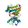


 PDBj
PDBj


 集合体
集合体


 分子量: 417.417 Da / 分子数: 1 / 由来タイプ: 合成 / 式: C22H19N5O4
 分子量: 18.015 Da / 分子数: 86 / 由来タイプ: 天然 / 式: H2O
 試料調製
試料調製 解析
解析