 Movie
Movie Controller

[English] 日本語
 Yorodumi
- PDB-4fd9: Crystal structure of the third beta-gamma-crystallin domain of Cr... -
+ Open data

- Basic information
| Entry | Database: PDB / ID: 4fd9 | ||||||
|---|---|---|---|---|---|---|---|
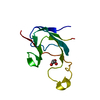
| Title | Crystal structure of the third beta-gamma-crystallin domain of Crybg3 (betagamma-crystallin domain-containing protein 3) from Mus musculus | ||||||
 Components Components | Beta/gamma crystallin domain-containing protein 3 | ||||||
 Keywords Keywords |  STRUCTURAL PROTEIN / Non-lens vertebrate beta-gamma-crystallin / Brain STRUCTURAL PROTEIN / Non-lens vertebrate beta-gamma-crystallin / Brain | ||||||
| Function / homology |  Function and homology information Function and homology informationstructural constituent of eye lens / lens development in camera-type eye / protein kinase A binding / visual perception / carbohydrate binding / protein-containing complexSimilarity search - Function | ||||||
| Biological species |  Mus musculus (house mouse) Mus musculus (house mouse) | ||||||
| Method | X-RAY DIFFRACTION / MOLECULAR REPLACEMENT / Resolution: 1.86 Å | ||||||
 Authors Authors | Aravind, P. / Srivastava, S.S. / Sankaranarayanan, R. | ||||||
 Citation Citation | Journal: Biochemistry / Year: 2012 Title: Aggregation-prone near-native intermediate formation during unfolding of a structurally similar nonlenticular beta/gamma-crystallin domain Authors: Rajanikanth, V. / Srivastava, S.S. / Singh, A.K. / Rajyalakshmi, M. / Chandra, K. / Aravind, P. / Sankaranarayanan, R. / Sharma, Y. | ||||||
| History |
|
- Structure visualization
| Structure viewer | Molecule: MolmilJmol/JSmol |
|---|
- Downloads & links
-Download
| PDBx/mmCIF format | 4fd9.cif.gz | 52.1 KB | Display | PDBx/mmCIF format |
|---|---|---|---|---|
| PDB format | pdb4fd9.ent.gz | 37.8 KB | Display | PDB format |
| PDBx/mmJSON format | 4fd9.json.gz | Tree view | PDBx/mmJSON format | |
| Others |  Other downloads Other downloads |
-Validation report
| Arichive directory | https://data.pdbj.org/pub/pdb/validation_reports/fd/4fd9ftp://data.pdbj.org/pub/pdb/validation_reports/fd/4fd9 | HTTPS FTP |
|---|
-Related structure data
| Related structure data | |
|---|---|
| Similar structure data |


-Links
 PDBj
PDBj


- Assembly
| Deposited unit | 
| ||||||||
|---|---|---|---|---|---|---|---|---|---|
| 1 | 
| ||||||||
| 2 | 
| ||||||||
| Unit cell |
|
-Components
| #1: Protein | Mass: 10434.805 Da / Num. of mol.: 2 / Fragment: beta-gamma-crystallin domain Source method: isolated from a genetically manipulated source Source: (gene. exp.) Mus musculus (house mouse) / Strain: C57BL/6 / Tissue: Brain / Gene: Crybg3 / Plasmid: pET21a / Production host:  Escherichia coli (E. coli) / Strain (production host): BL21(DE3) / References: UniProt: Q80W49 Escherichia coli (E. coli) / Strain (production host): BL21(DE3) / References: UniProt: Q80W49#2: Water | ChemComp-HOH / | Water Mass: 18.015 Da / Num. of mol.: 210 / Source method: isolated from a natural source / Formula: H2O Mass: 18.015 Da / Num. of mol.: 210 / Source method: isolated from a natural source / Formula: H2O |
|---|
-Experimental details
-Experiment
| Experiment | Method: X-RAY DIFFRACTION / Number of used crystals: 1 |
|---|
- Sample preparation
| Crystal | Density Matthews: 1.99 Å3/Da / Density % sol: 38.25 % |
|---|---|
| Crystal grow | Temperature: 277 K / Method: vapor diffusion, hanging drop / pH: 7.5 Details: 20-25% PEG 4K, 0.1M Na Hepes, 10% isopropanol , pH 7.5, VAPOR DIFFUSION, HANGING DROP, temperature 277K |
-Data collection
| Diffraction | Mean temperature: 100 K | |||||||||||||||
|---|---|---|---|---|---|---|---|---|---|---|---|---|---|---|---|---|
| Diffraction source | Source: ROTATING ANODE / Type: RIGAKU RUH3R / Wavelength: 1.5418 Å | |||||||||||||||
| Detector | Type: MAR scanner 345 mm plate / Detector: IMAGE PLATE / Date: Jul 26, 2007 | |||||||||||||||
| Radiation | Protocol: SINGLE WAVELENGTH / Monochromatic (M) / Laue (L): M / Scattering type: x-ray | |||||||||||||||
| Radiation wavelength | Wavelength: 1.5418 Å / Relative weight: 1 | |||||||||||||||
| Reflection twin |
| |||||||||||||||
| Reflection | Resolution: 1.86→25 Å / Num. obs: 13629 / Redundancy: 5.7 % / Rmerge(I) obs: 0.037 | |||||||||||||||
| Reflection shell | Resolution: 1.86→1.93 Å / Redundancy: 5 % / Rmerge(I) obs: 0.2 / Num. unique all: 1167 |
- Processing
| Software |
| |||||||||||||||||||||||||||||||||||||||||||||
|---|---|---|---|---|---|---|---|---|---|---|---|---|---|---|---|---|---|---|---|---|---|---|---|---|---|---|---|---|---|---|---|---|---|---|---|---|---|---|---|---|---|---|---|---|---|---|
| Refinement | Method to determine structure: MOLECULAR REPLACEMENT / Resolution: 1.86→22.78 Å / Cor.coef. Fo:Fc: 0.97 / Cor.coef. Fo:Fc free: 0.944 / SU B: 2.774 / SU ML: 0.087 / Cross valid method: THROUGHOUT / ESU R: 0.029 / ESU R Free: 0.026 / Stereochemistry target values: MAXIMUM LIKELIHOOD / Details: HYDROGENS HAVE BEEN USED IF PRESENT IN THE INPUT
| |||||||||||||||||||||||||||||||||||||||||||||
| Solvent computation | Ion probe radii: 0.8 Å / Shrinkage radii: 0.8 Å / VDW probe radii: 1.2 Å / Solvent model: MASK | |||||||||||||||||||||||||||||||||||||||||||||
| Displacement parameters | Biso mean: 24.189 Å2
| |||||||||||||||||||||||||||||||||||||||||||||
| Refinement step | Cycle: LAST / Resolution: 1.86→22.78 Å
| |||||||||||||||||||||||||||||||||||||||||||||
| Refine LS restraints |
| |||||||||||||||||||||||||||||||||||||||||||||
| LS refinement shell | Resolution: 1.865→1.913 Å / Total num. of bins used: 20
|
